Scientists created miniature brains in the lab that formed intricate networks and produced brain waves similar to those fired by the developing brain of a preterm human baby, according to a new study.
The idea of growing miniature brains in the lab isn't new; researchers have been doing so for nearly a decade. But most studies have used these mini brains or "organoids" to study large-scale structure.
For example, one group developed mini brains that could grow blood vessels, Live Science previously reported. Another group exposed mini brains to the Zika virus to understand how it may lead to abnormally small heads, or microcephaly.
Related: 11 Body Parts Grown in the Lab
But in conditions such as autism, schizophrenia, bipolar disorder and even depression, "the brain is intact and the problem relies on the operations of the network," said study senior author Alysson Muotri, an associate professor the Department of Cellular and Molecular Medicine and the director of the Stem Cell Program at the University of California, San Diego. This is the first time lab-grown brains have formed intricate networks of neurons that produced strong brain waves.
To do this, Muotri and his team harvested human stem cells — which can morph into any cell type given the right instructions — derived from people's skin and blood. The researchers exposed these stem cells to chemical instructions that would turn the cells into brain cells.
For the most part, these cells formed neural progenitor cells, brain-specific cells that can proliferate and give rise to many types of brain cells. After two to five months in a lab dish, these progenitor cells form glutamatergic neurons, brain cells that are "excitatory," or those that propagate information.
After about four months, the mini brains stopped making excitatory neurons and began making astrocytes. These brain cells help shape synapses, the gaps between brain cells where neurotransmitters, or brain chemicals, pass information. Finally, the progenitor cells began making inhibitory neurons, which quench brain activity, or stop neurons from passing information. That's when "the activity starts to become more complex, because now we [are] balancing excitation and inhibition," Muotri said.
While the cells were dividing and differentiating, they eventually began to "self-organize into something that resembles the human cortex," Muotri said. The cortex is the outer layer of the brain, which plays an important role in consciousness.
The "mini brains" don't, in fact, look like miniature versions of human brains. Rather, they are white, spherical blobs that float in the reddish soup in which they're grown, Muotri said. They grew up to only 0.2 inches (0.5 centimeters) in diameter, but their neural networks continued to evolve for nine to 10 months before stopping, he said.
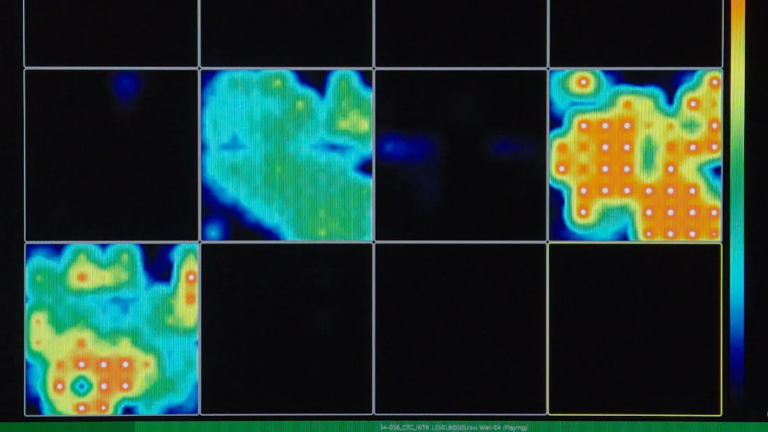
Throughout the growth of the mini brains, the team used a set of tiny electrodes that connect to neurons to measure brain activity. The researchers found that at around two months, the neurons in the mini brains began to fire sporadic signals, all at the same frequency. After a couple more months of development, the brains fired signals at different frequencies and more regularly, indicating more-complex brain activity, Muotri said.
While previous studies have shown that mini, lab-produced brains could produce brain cell firing, researchers reported them firing around say 3,000 times per minute, Muotri said. In this study, however, the neurons fired close to 300,000 times per minute, which is "closer to the human brain," he said.
The team then used a machine-learning algorithm to compare the brain activity of these mini brains to that of preterm human babies. The researchers trained their program to learn the brain waves recorded from 39 premature babies between 6 and 9-and-a-half months old.
The scientists then fed the brain wave patterns from the mini brains into the algorithm and found that after 25 weeks of mini brain development, it could no longer distinguish the data coming from the human brain from that deriving from the lab-grown brain. "It gets confused and gives the same age to both of them," which suggests that the mini brains and the human brains were growing and developing similarly, Muotri said.
This study shows "very nicely that you can make this reproducible experimental systems where you can address processes which are so fundamental for the development of a human being", said Dr. Thomas Hartung, the director of the Johns Hopkins Center for Alternatives to Animal Testing who has also worked on developing mini-brains in the lab but who was not a part of the study.
The "inaccessibility of the embryonic brain is one of the reasons why these models are offering something different," he said. "But it also means you have very limited opportunities to say its the real thing." While the EEG signals are similar to that of pre-term babies, they're slightly off in timing, he added.
While a human embryo is connected to the mother and thus receives signals from the outside, these lab-grown brains aren't connected to anything. "These cells have no input or no output they cannot recognize anything happening in the world," Hartung said. So they are "definitely not" conscious.
That's what most scientists would agree on, but "it's hard to say," Muotri said. "We neuroscientists don't even agree [on] what are the measurements that one can do to actually probe to see if they're conscious or not."
The human brain sends its signals to help us interact with our environment. For example, we look at a bug, the eyes send signals to brain cells, which signal to each other and let us know that we are seeing a bug.
So, why are these lab-grown brains sending signals? What could they possibly be talking about? "That's a question we don't know, because the embryonic brain is really a black box," Muotri said. It seems that most of the signals in these early stages involve instructions to "self-wire," or connect to each other, he said.
In any case, he said he hopes studies like this will help us understand how early brain wiring gives rise to our complex brains, and what happens when that wiring goes awry.
Muotri and his team said they now hope to stimulate the brain organoids further to see if they can develop beyond nine to 10 months. The researchers would also like to model brain disorders, for instance by creating brain organoids with cells taken from children with autism, to understand how their brain networks develop.
The findings were published today (Aug. 29) in the journal Cell Stem Cell.
- Top 3 Techniques for Creating Organs in the Lab
- 7 Ways to Trick Your Brain
- 3D Images: Exploring the Human Brain
Originally published on Live Science.

