It's quick and easy to access Live Science Plus, simply enter your email below. We'll send you a confirmation and sign you up for our daily newsletter, keeping you up to date with the latest science news.
Incredible 3D images of over 13,000 vertebrates — representing half of the world's described genera — have been created as part of a project to make museum specimens available to all.
From spine-tailed mice (Acomys species) to rare rim rock crowned snakes (Tantilla oolitica), natural collections from museums around the world are being added to openVertebrate (oVert) — a five-year project funded by the National Science Foundation creating a database of computed tomography (CT) scans of specimens.
CT scans combine multiple X-ray images taken from different angles around the body to create detailed cross-sectional images, enabling scientists to peer through the exterior of animals without damaging the specimens. This gives them valuable insight into skeletal structures.
Some specimens were even stained during scanning so researchers could view soft tissue structures, revealing stomach contents, eggs, parasites and organs.
Using various methods, researchers can reconstruct museum specimens as digital 3D models.
(Image credit: openVertebrate)
As of November 2023, 13,000 specimens across 18 U.S. institutions have been scanned.
The project team has released a selection of images from the project showing creatures in remarkable detail. These include a CT scan of the final meal of a hognose snake (Heterodon platirhinos), the prickly spines of a four-toed hedgehog (Atelerix albiventris) and a black-bellied fruit bat (Melonycteris melanops).
Sign up for the Live Science daily newsletter now
Get the world’s most fascinating discoveries delivered straight to your inbox.
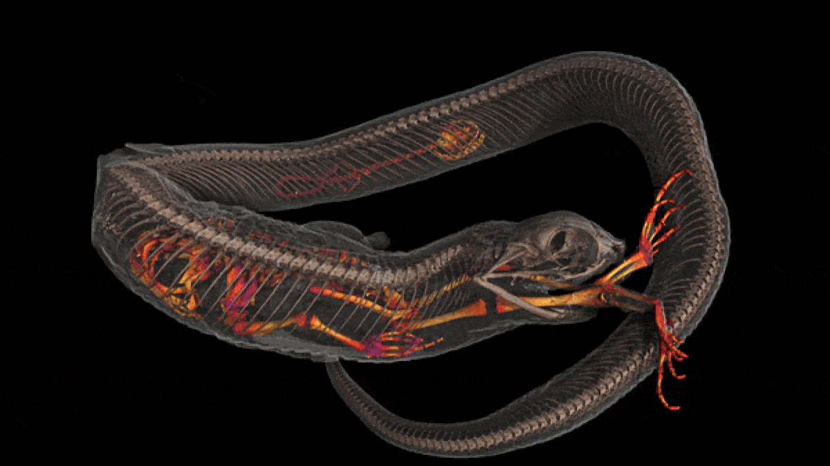
A computed tomography scan showing the final two meals of an Eastern hognose snake (Heterodon platirhinos): a salamander and a toad.
(Image credit: Credit: Edward Stanley and David Blackburn, Florida Museum of Natural History)
When first created, museums held rare specimens as part of private collections of wealthy individuals. Now, museums are open to the public, but some collections remain concealed.
"Access to these collections is often limited; cost of travel, space restrictions, fragility of specimens, all can prevent people from working with these samples," Edward L. Stanley, an author and Director of the Digital Imaging Division at the Florida Museum of Natural History, told Live Science in an email.
CT scan of the skeletal structure of a black-bellied fruit bat (Melonycteris melanops).
(Image credit: openVertebrate)
CT scan of an oriental fire-bellied toad, showing mineralization in the skin.
(Image credit: openVertebrate)
CT scan showing the skeletal structure of a four-toed hedgehog (Atelerix albiventris).
(Image credit: openVertebrate)
CT scan of a cardinal soldierfish (Plectrypops retrospinosus).
(Image credit: openVertebrate)
CT scan of a species of toad from the genus, Alytes, showing its heart.
Stanley is an author of a study published Mar 6. in the journal BioScience that provides a summary of the project to date.
Studying specimens often involves dissections and chemical testing, which can damage specimens, further limiting accessibility. "Museums have to strike a balance between using their specimens and using them up, which often means that rarest and most important specimens are often the hardest to access", Stanley said.
"The digital data from CT scanning provides a fast and low-risk way for museums to make even their most delicate, rare or irreplaceable specimens available to anyone, anywhere in the world," Stanley added.

 Live Science Plus