Spangled Beetle Eye and Retinal 'Fireworks' Dazzle in Nikon Photo Contest

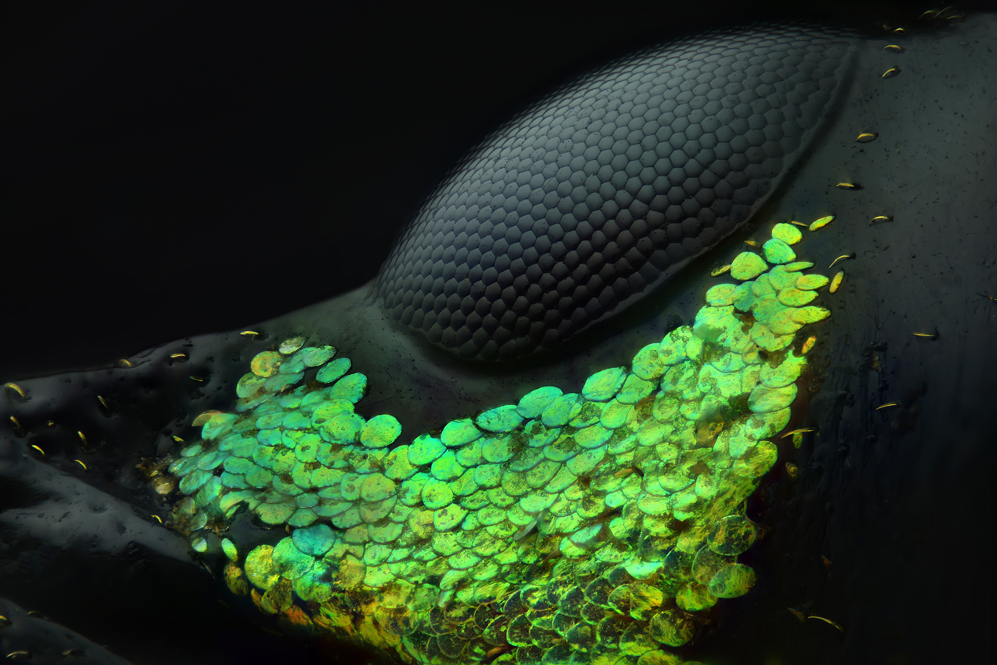
A beetle eye surrounded by glittery emerald-green scales took the top prize in this year's Nikon Small World microphotography challenge.
The annual contest, now in its 44th year, celebrates stunning images of objects too small to be seen without a microscope. This year, winning photographers turned their magnifying camera lenses toward such wee marvels as a spider embryo, filaments in a peacock feather and illuminated blood vessels in the oviduct of a mouse.
First prize went to photographer Yousef Al Habshi of Abu Dhabi, United Arab Emirates, for his spectacular image showing the pitch-black compound eye of an Asian red palm weevil (Metapocyrtus subquadrulifer). In the photo, the beetle's dark eye is ringed by tiny yellow hairs and an expanse of glowing scales in shades of brilliant green, which was captured by stacking dozens of images, Nikon representatives said in a statement released Oct. 11. [Photos: Peer at Glittering Insect Eyes and Glowing Barnacle Hairs in Prizewinning Photos]
Capturing close-up photos of a weevil's head is challenging, to say the least, as the insect's entire body typically measures less than 0.4 inches (11 millimeters). Al Habshi stacked 128 micrographs to achieve the remarkable color and texture details in his image, which balanced the deep black of the beetle's eye and skin with the brilliant green of the scales around it, he explained in the statement.
"Not all people appreciate small species, particularly insects," he said. "Through photomicrography we can find a whole new, beautiful world which hasn't been seen before."
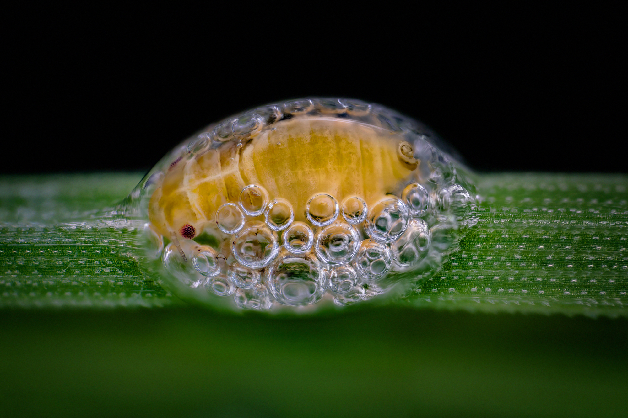
A colorful fluorescent cluster of spore-producing structures in a fern earned the second place spot for Rogelio Moreno Gill, a photographer in Panama City, Panama; while Naperville, Illinois, photographer Saulius Gugis nabbed third place with a photo of a bubble-blowing spittlebug nymph huddled inside its bubble home on a leaf.
Other standout photos include a spider embryo, its baby legs wrapped tightly around its tiny body; the central region of a human retina, illuminated like a fireworks display; and the incredibly intricate, frond-like structures inside a single human teardrop.
Get the world’s most fascinating discoveries delivered straight to your inbox.
"Every year, we continue to be astounded by the winning images," Nikon Instruments representative Eric Flem said in the statement. "Imaging and microscope technologies continue to develop and evolve to allow artists and scientists to capture scientific moments with remarkable clarity," Flem said.
You can see all of the winners, honorable mentions and images of distinction — 95 in all — on the Nikon Small World website.
Originally published on Live Science.

Mindy Weisberger is a science journalist and author of "Rise of the Zombie Bugs: The Surprising Science of Parasitic Mind-Control" (Hopkins Press). She formerly edited for Scholastic and was a channel editor and senior writer for Live Science. She has reported on general science, covering climate change, paleontology, biology and space. Mindy studied film at Columbia University; prior to LS, she produced, wrote and directed media for the American Museum of Natural History in NYC. Her videos about dinosaurs, astrophysics, biodiversity and evolution appear in museums and science centers worldwide, earning awards such as the CINE Golden Eagle and the Communicator Award of Excellence. Her writing has also appeared in Scientific American, The Washington Post, How It Works Magazine and CNN.
 Live Science Plus


