
 Live Science Plus


Insect Tangled Up in Blue in Amazing Microphotograph

By
Nina Sen
published
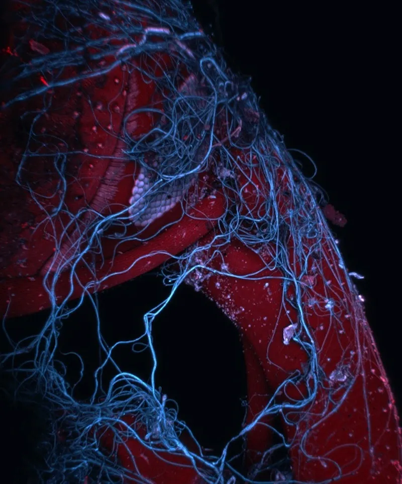
This microphotograph of an insect trapped in a spider web by Mark A. Sanders from University Imaging Centers, University of Minnesota placed ninth at Nikon's 2013 Small World Photomicrography competition.
(Image credit: Mark A. Sanders / University Imaging Centers, University of Minnesota)
This amazing image is actually of an insect wrapped up in a spider web took ninth place at Nikon's 2013 Small World Photomicrography competition.
Mark A. Sanders from University Imaging Centers, University of Minnesota in Minneapolis, Minn. submitted the photo, which was created from stacked images magnified 85 times. He used autofluorescence and confocal optical imaging technique to capture the false-color photo.

