
 Live Science Plus


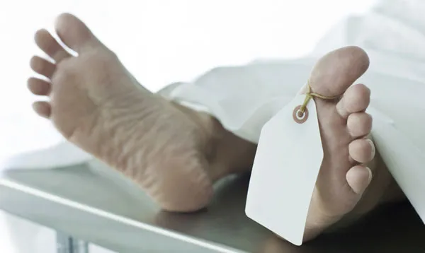
What Exactly Do They Do During an Autopsy?
What most of us know of autopsies comes from popular television crime dramas, with their super-sleuth forensics teams and equipment so cutting-edge it borders on science fiction. We're spoiled by the convenient swiftness with which good-looking medical examiners obtain results, and the sheer volume of detailed information they get from the tiniest clues — so much so that some now decry a trend of unrealistic evidentiary expectations among jurors, dubbing it the "CSI Effect."
Perhaps it's time we looked at what actually goes on during an autopsy.
An autopsy is an examination of a dead body to determine cause of death , the effects or indications of disease or, in some cases, to identity the dead person. Forensic pathologists — physicians trained in the study of diseases and abnormalities — perform autopsies with the assistance of autopsy technicians (sometimes called "dieners," from the German for “helper”) and autopsy photographers.

The type of autopsy most familiar from television and movies is the kind legally ordered by the state to resolve violent, suspicious or sudden deaths. However, autopsies are also performed for disease research and medical training.
Before conducting an autopsy, investigators gather all the information they can about the subject and the events leading to his or her demise, consulting medical records, doctors and family members and examining the location and circumstances of death.
External examination
The autopsy begins with a careful inspection of the body. This can help establish identity, locate evidence or suggest a cause of death. The pathologists weigh and measure the body, noting the subject's clothing, valuables and characteristics such as eye color, hair color and length, ethnicity, sex and age.
Get the world’s most fascinating discoveries delivered straight to your inbox.
Removing the subject's clothes, they then examine the body, searching for gunpowder residue, paint flakes or other deposits, identifying marks such as scars or tattoos, or injuries. X-rays are sometimes used to reveal bone abnormalities and the locations of bullets or other objects, and ultraviolet light can help detect certain residues. Pathologists may also take samples of hair and nails at this time.
Throughout the autopsy, the pathologist records everything on a body diagram and in recorded verbal notes.
Internal examination
If a complete internal examination is called for, the pathologist removes and dissects the chest, abdominal and pelvic organs, and (if necessary) the brain. It is unusual to examine the face, arms, hands or legs internally. The cuts into the body produce little blood because without a beating heart the only blood pressure comes from gravity.
Prior to cutting, the torso is placed on a rubber block, extending the body's arch and providing greater access to the chest and abdomen. If a brain autopsy is also planned, this block will be moved to support the head once the torso work is complete.
The pathologist begins the chest and abdomen autopsy by making a Y-shaped incision, the two arms of the Y running from each shoulder joint,to meet at mid-chest and the stem of the Y running down to the pubic region. This is one of the aspects of autopsies that movies and television shows get wrong, according to Dr. Ed Uthman, a Texas pathologist who has written a screenwriter's guide to autopsies.
"The most common error is making the trunk incision wrong," Uthman said. "On women, the two arms of the Y are supposed to curve around under the breasts , but in films, they invariably show them straight and above the breasts."
"Also, in both sexes, they make the arms of the Y too short; they actually need to extend all the way up to each shoulder joint," Uthman said.
The next step is to examine the organs in situ (in place), which means removing the rib cage. Using a saw or a rib cutter (similar in appearance to a small pruning shear), the pathologists cut along the boundary between the ribs and the cartilage connected to the breastbone. Alternatively, they might cut the sides of the chest cavity, leaving the ribs attached to the breastbone and removing the entire frontal ribcage as one chest plate.
The abdominal examination begins with a pathologist freeing the intestines by cutting along the attachment tissue with scissors or a scalpel.
If a brain autopsy is called for, the pathologist will make a cut across the crown of the head, from the bony bump behind one ear to the bump behind the other. He or she will then open the cranium using a special saw that cuts bone but leaves soft tissue unharmed.
Once each organ has been examined within the body, it is removed, weighed and examined in further detail. Sometimes organs are removed individually, a procedure referred to as the Virchow technique; other times, they are removed as a connected group, via the Rokitansky technique.
"I like the Rokitansky myself, because it frees up the body earlier, so the diener can get to work with the closing and cleanup," Uthman said.
Organs, especially the brain, are sometimes placed in formalin for days or even weeks before the dissection is conducted. Formalin preserves organs while also granting them greater firmness, allowing for neater and more accurate dissections.
In particular, brain tissue benefits from fixation in formalin because its natural texture resembles soft gelatin or firm tofu. According to Uthman, a few weeks in the fixative lend the brain "the consistency and firmness of a ripe avocado." Once removed, the lungs may also be inflated with fixative.
Tissue samples are taken from the organs, some of which may be also be sectioned, and stomach contents are frequently tested. Pathologists and lab technicians also test bodily fluids — urine, blood, vitreous gel from the eyes, or bile from the gallbladder — for drugs, infection, chemical composition or genetic factors, depending on the purpose of the autopsy.
Pathologists will preserve parts of any organs they dissect, particularly if they find something unusual or abnormal.
Reconstituting the body
Following examination, the organs are either returned to the body (minus the pieces preserved for future work or evidence) or cremated, in accordance with the law and the family's wishes. The breastbone and ribs are also usually put back.
Prior to being sewn shut with the characteristic "baseball stitch," the body is lined with cotton wool or a similar material. If the organs are to be returned to the body, they are first placed in bags to prevent leakage. The body is then sewn shut, washed and prepared for the funeral director.
Bodies that have undergone autopsy are still able to have open-casket funerals, even in the case of brain autopsy: A casket pillow will hide the cranial cut.
Follow Life's Little Mysteries on Twitter @nattyover or Life's Little Mysteries @llmysteries. We're also on Facebook & Google+.