 Live Science Plus


You are now subscribed
Your newsletter sign-up was successful
Want to add more newsletters?

Delivered Daily
Daily Newsletter
Sign up for the latest discoveries, groundbreaking research and fascinating breakthroughs that impact you and the wider world direct to your inbox.

Once a week
Life's Little Mysteries
Feed your curiosity with an exclusive mystery every week, solved with science and delivered direct to your inbox before it's seen anywhere else.

Once a week
How It Works
Sign up to our free science & technology newsletter for your weekly fix of fascinating articles, quick quizzes, amazing images, and more

Delivered daily
Space.com Newsletter
Breaking space news, the latest updates on rocket launches, skywatching events and more!

Once a month
Watch This Space
Sign up to our monthly entertainment newsletter to keep up with all our coverage of the latest sci-fi and space movies, tv shows, games and books.

Once a week
Night Sky This Week
Discover this week's must-see night sky events, moon phases, and stunning astrophotos. Sign up for our skywatching newsletter and explore the universe with us!
Join the club
Get full access to premium articles, exclusive features and a growing list of member rewards.
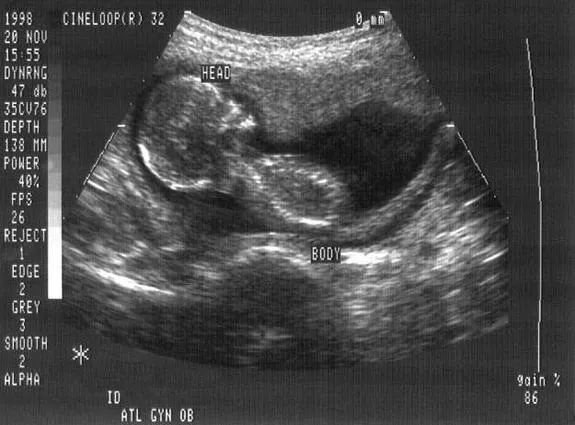
For most women today, it's hard to imagine going through a pregnancy without having an ultrasound. But these iconic black-and-white images of a developing fetus, generated by the reflection of high frequency sound waves, have only been around since the mid-1950s.
A new book explores the history of ultrasounds, in both their technical and social dimensions. In "Imaging and Imagining the Fetus: The Development of Obstetric Ultrasound" (The Johns Hopkins University Press, 2013), authors Malcolm Nicolson, a history of medicine professor at the University of Glasgow in Scotland, and engineer John Fleming look at how ultrasounds came into wide use, and why their images lie at the crossroads of several hotly debated issues today.
Ultrasound was first used for clinical purposes in 1956 in Glasgow. Obstetrician Ian Donald and engineer Tom Brown developed the first prototype systems based on an instrument used to detect industrial flaws in ships.
They perfected its clinical use, and by the end of the 1950s, ultrasound was routinely used in Glasgow hospitals, Nicolson said. But it didn’t really take off in British hospitals until the 1970s, and it was well into the 1970s before it became widely used in American hospitals, he said. [Blossoming Body: 8 Odd Changes That Happen During Pregnancy]
By the end of the 20th century, ultrasound imaging had become routine in maternity clinics throughout the developed world. The technology has undergone extensive development over the past 20 years, Nicolson told LiveScience, but "has probably reached more or less the pinnacle of its acuity."
How does it work?
Ultrasound imaging involves bouncing "ultrasonic" sound waves — above the audible range of human hearing — at body structures or tissues, and detecting the echoes that bounce back.
—Abortion laws by state: https://reproductiverights.org/maps/abortion-laws-by-state/
—For questions about legal rights and self-managed abortion: www.reprolegalhelpline.org
—To find an abortion clinic in the US: www.ineedanA.com
—Miscarriage & Abortion Hotline operated by doctors who can offer expert medical advice: Available online or at 833-246-2632
—To find practical support accessing abortion: www.apiarycollective.org
Obstetric ultrasonography is used to image a human fetus inside its mother's womb. It's used to confirm a pregnancy, to identify the sex and number of fetuses and to detect fetal abnormalities such as microcephaly (an abnormally small head), absence of kidneys, and spinal problems.
During a scan, ultrasound waves are aimed at a pregnant women's abdomen. Based on the angle of the beam, and the time it takes for echoes to return, an image of body structures inside the fetus can be generated.
Early in the use of fetal ultrasound, clinicians could only detect the baby's head, Nicolson said. "But gradually, with developing expertise, they could discern fine structures in the fetus," he said.
Is it safe?
One of the main advantages of ultrasound is that it's noninvasive. The procedure has been safely performed on millions of pregnant women. Concerns about its safety have periodically surfaced, but Nicolson said he thinks these stem more from anxiety over the role of technology in pregnancy than from evidence of harm.
"We can be confident that at the levels currently used for clinical investigation, ultrasound is safe. No pattern of damage has been found," Nicolson said.
However, at high power, ultrasound waves are able to damage human tissue. Researchers don't know exactly at what level this occurs, Nicolson said, adding that testing the threshold at which it becomes dangerous in humans would be unethical.
He said that ultrasound scans should only be done for clinically justified reasons. For example, so-called "bonding scans," images taken purely for commemorative purposes, unnecessarily expose a fetus to the high-energy sound waves, Nicolson said.
What's the emotional impact?
Ultrasound has enjoyed an enthusiastic reception by pregnant women. In addition to revealing the baby's health, the images themselves provide a keepsake. "Overwhelmingly, pregnant women expect to be scanned, and are moved and excited by seeing the fetus," Nicolson said — especially if the baby moves. In fact, Nicolson said, some women report not feeling pregnant until they've seen the ultrasound image.
Seeing a developing fetus has a humanizing effect, too. Donald, the physician who helped develop the technology, was a devout High Anglican, and knew the images carried moral significance for women contemplating having an abortion.
Are there social implications?
Ultrasound images sometimes play a role in decisions to maintain or terminate a pregnancy. Anti-abortion proponents take ultrasound images as proof that a fetus is fully alive and therefore should not be aborted.
On the other hand, ultrasound can be used to diagnose potentially fatal or debilitating abnormalities in the fetus, which can encourage termination of the pregnancy.
In some East Asian countries, ultrasound is used to detect the sex of the baby expressly so that a fetus of less desirable sex (usually female) can be aborted, Nicolson said. He called the practice "unfortunate and worrying."
Still, anecdotal evidence suggests that if pregnant women see images of a fetus — especially their own — they are less likely to terminate a pregnancy, Nicolson said.
"The realm of human reproduction is an area that is contested, and bound to be emotive," Nicolson said.
Follow Tanya Lewis on Twitter and Google+. Follow us @livescience, Facebook & Google+. Original article on Live Science.
Editor's note: This article was updated on August 3, 2022 by Live Science contributor Alice Ball following the Supreme Court's decision to overturn Roe v. Wade on June 24, 2022. This decision eliminated the constitutional right to abortion that was established by the 1973 court case and later affirmed by a 1992 case called Planned Parenthood of Southeastern Pennsylvania v. Casey.

