


Lab-made mouse embryos grew brains and beating hearts, just like the real thing
The embryos survived for 8.5 days.
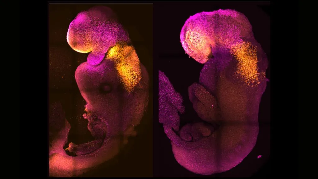
Scientists coaxed mouse stem cells to grow into synthetic embryos that began developing hearts and brains, just like the real thing.
The lab-made embryos, crafted without any eggs or sperm and incubated in a device that resembles a fast-spinning Ferris wheel full of tiny glass vials, survived for 8.5 days. That's nearly half the length of a typical mouse pregnancy. In that time, a yolk sac developed around the embryos to supply nutrition, and the embryos themselves developed digestive tracts; neural tubes, or the beginnings of the central nervous system; beating hearts; and brains with well-defined subsections, including the forebrain and midbrain, the scientists reported in a study published Thursday (Aug. 25) in the journal Nature.
"This has been the dream of our community for years and [a] major focus of our work for a decade, and finally, we've done it," senior study author Magdalena Zernicka-Goetz, a developmental and stem-cell biologist with labs at the University of Cambridge, UK, and the California Institute of Technology in Pasadena, said in a statement.
The new work produced very similar results as an earlier study, published Aug. 1 in the journal Cell, which was led by Jacob Hanna, an embryonic stem cell biologist at the Weizmann Institute of Science in Israel and co-author of the new Nature paper. In their recent Cell study, Hanna's team used different starting stem cells but the same incubator to culture synthetic mouse embryos for 8.5 days. Those embryos also grew digestive tracts, beating hearts, and tiny, wrinkled brains before ultimately dying, Live Science previously reported.
Related: 'First complete models' of a human embryo made in the lab
Although the two recent studies produced similar embryos, the experiments started out slightly differently. In the Cell study, the researchers started by coaxing mouse stem cells into a naive state from which they could morph into any cell type, such as heart, brain or gut cells. From there, the team divided these naive cells into three groups. In one group, they switched on genes to form the placenta, and in another group, they switched on genes to make the yolk sac. The last group they left alone to develop into embryos.
Zernicka-Goetz's research group, on the other hand, began with three mouse stem cell types, rather than starting with only naive cells. One type of stem cell gave rise to the embryo, while the other two morphed into the placental tissues and yolk sac. Throughout the experiment, they observed how these three stem cell types interacted, exchanging chemical messages and physically butting up against each other in the glass vials.
Get the world’s most fascinating discoveries delivered straight to your inbox.
Studying such exchanges could give hints as to how the earliest stages of embryonic development unfold in humans — and what happens when things go awry.
"This period of human life is so mysterious, so to be able to see how it happens in a dish — to have access to these individual stem cells, to understand why so many pregnancies fail and how we might be able to prevent that from happening — is quite special," Zernicka-Goetz said. "We looked at the dialogue that has to happen between the different types of stem cell at that time — we've shown how it occurs and how it can go wrong."
RELATED STORIES
In both the Cell and Nature studies, the resulting synthetic embryos closely resembled natural embryos, albeit with some slight differences and defects in how the tissues self-organized. However, in both experiments, a very low proportion of the stem cells actually gave rise to embryos, suggesting that the efficiency of both systems could be improved. In addition, neither set of synthetic embryos survived to the ninth day of development — an obstacle that would need to be overcome in follow-up studies.
"The reason for the block in further development is unclear but might relate to the defects in the formation of some of the placental cell types that the authors report," James Briscoe, a principal group leader and assistant research director at the Francis Crick Institute in the U.K. who was not involved in either study, told the Science Media Centre, a U.K.-based press office that works with researchers, journalists and policymakers to disseminate accurate scientific information.
The research also raises ethical questions about if and how such technology might be applied to human cells in the future.
Originally published on Live Science.

Nicoletta Lanese is the health channel editor at Live Science and was previously a news editor and staff writer at the site. She is a recipient of the 2026 AHCJ International Health Study Fellowship, with a project focused on antibiotic stewardship practices in Japan and the U.S. They hold a graduate certificate in science communication from UC Santa Cruz and degrees in neuroscience and dance from the University of Florida. Beyond Live Science, Lanese's work has appeared in The Scientist, Science News, the Mercury News, Mongabay and Stanford Medicine Magazine, among other outlets. Based in NYC, she also remains involved in dance and performs in local choreographers' work.