It's quick and easy to access Live Science Plus, simply enter your email below. We'll send you a confirmation and sign you up for our daily newsletter, keeping you up to date with the latest science news.
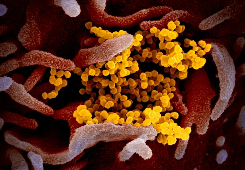
This scanning electron microscope image shows SARS-CoV-2 (yellow) among human cells (pink). This virus was isolated from a patient in the U.S. (Color has been added to the image to better show the virus and its environment.)
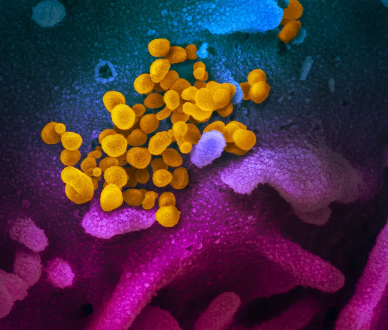
On Thursday (Feb. 13), the Rocky Mountain Laboratories (RML) at the National Institute of Allergy and Infectious Diseases revealed some of the first images of SARS-CoV-2, the new coronavirus that has sickened over 60,000 people and killed another 1,370 in the outbreak that began in Wuhan, China.
Viruses are teeny-tiny infectious blobs that are made up of either DNA or RNA wrapped up inside a protein coat. They are too small to be seen by a typical light microscope.
Researchers at RML imaged samples of the virus and cells taken from a U.S. patient infected with COVID-19 (the new name for the disease caused by SARS-CoV-2) using two different kinds of high-resolution microscopes — the scanning electron microscope and the transmission electron microscope. Both use a focused beam of electrons rather than a beam of light to image samples. (The color is added later to the images.)
This scanning electron microscope image shows the new coronavirus (yellow) among human cells (blue, pink and purple). (Color has been added to the image to better show the virus and its environment.)
(Image credit: NIAID-RML)
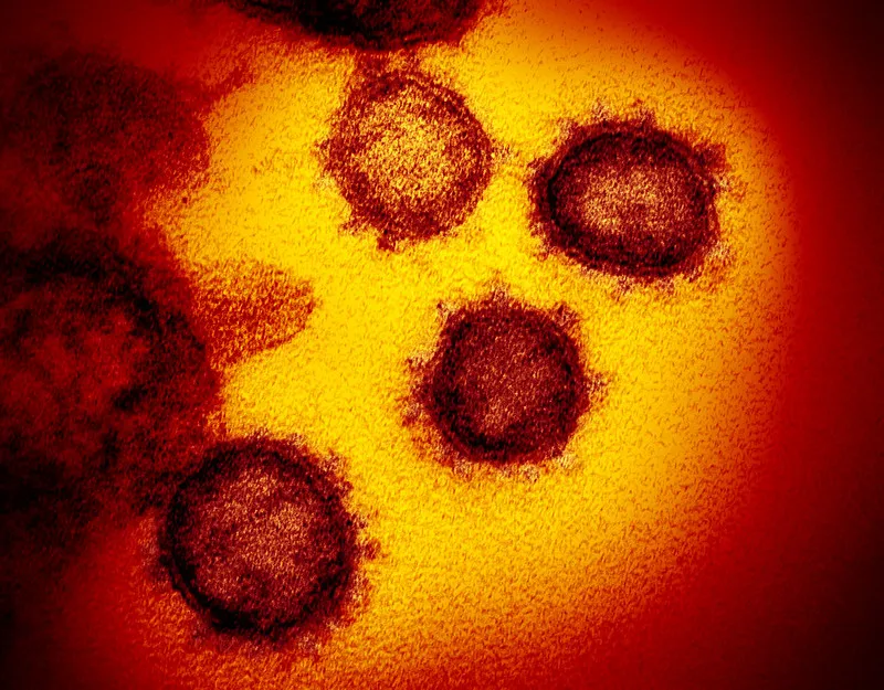
The SARS-COV-2 virus looks similar to the Middle East respiratory syndrome coronavirus (MERS-CoV), which emerged in 2012, and the severe acute respiratory syndrome coronavirus (SARS-CoV), which emerged in 2002, according to a statement.
That's because all three of these viruses are in the same family of "coronaviruses," which are named for their crown-like appearance (most apparent in the transmission electron image). The word "corona" in Latin means "crown."
This is a transmission electron microscope image showing the new coronavirus emerging from the surface of human cells.
Yasemin is a staff writer at Live Science, covering health, neuroscience and biology. Her work has appeared in Scientific American, Science and the San Jose Mercury News. She has a bachelor's degree in biomedical engineering from the University of Connecticut and a graduate certificate in science communication from the University of California, Santa Cruz.

 Live Science Plus