


Scientists in Switzerland have recently simulated a thin slice of a rat's brain in a computer. The effort relied on tens of thousands of experiments and billions of equations. The virtual brain slice captured some of the behavior exhibited by real brain cells. (Photo credits: Blue Brain Project, EPFL) [Read the full story on the digital rat brain]
Complicated setup

To recreate the brain, the team conducted many experiments on the neocortex of juvenile rats, cataloguing the interactions of many neurons. They also pored through all the existing literature for other experiments conducted on the rat's neocortex. From those studies, they derived general constraints and guiding principles for how neuronal networks are structured. Here, a view inside some of the neurons in the simulated brain.

Pruning neurons

To understand how the neurons interacted, they first created a network with 600 million connections — one for every point where a neuron would "touch" another. They then used a few guiding principles to prune these networks, resulting in a remaining 37 million connections. Here, some of the pruned networks shown in silico, with each type of neuronal shape or morphology painted in a different color.
Many things going on

Altogether, the construction captured many different types of cells, brain layers and structures. Here, a map shows how the model captures the interacting aspects of physiology and anatomy. Because the model includes so many potential variables, it must solve billions of equations just to simulate a blip in time in just this tiny portion of the rat's brain.
Get the world’s most fascinating discoveries delivered straight to your inbox.
Billions of equations

Once they had the connections, they created a virtual 3D volume in which brain cells were integrated. The model then produced billions of questions to solve for each 25 microseconds of time. Here, another view of the in silico brain slice, with different neuronal networks color coded.
Mimicking real life

While there's no way to know exactly how closely the juvenile rat brain mimics the real thing, there are some indications that the simulation is capturing neuronal behavior seen in the lab. For instance, the model showed evidence of a triplet pattern, in which three neurons fire in a tightly timed sequence. Here, an image of a virtual brain slice, with neurons captured using a stain that adheres to the Golgi apparatus in cells. The image looks very similar to those found in real brain slices using such staining methods.
Revealing new biology?

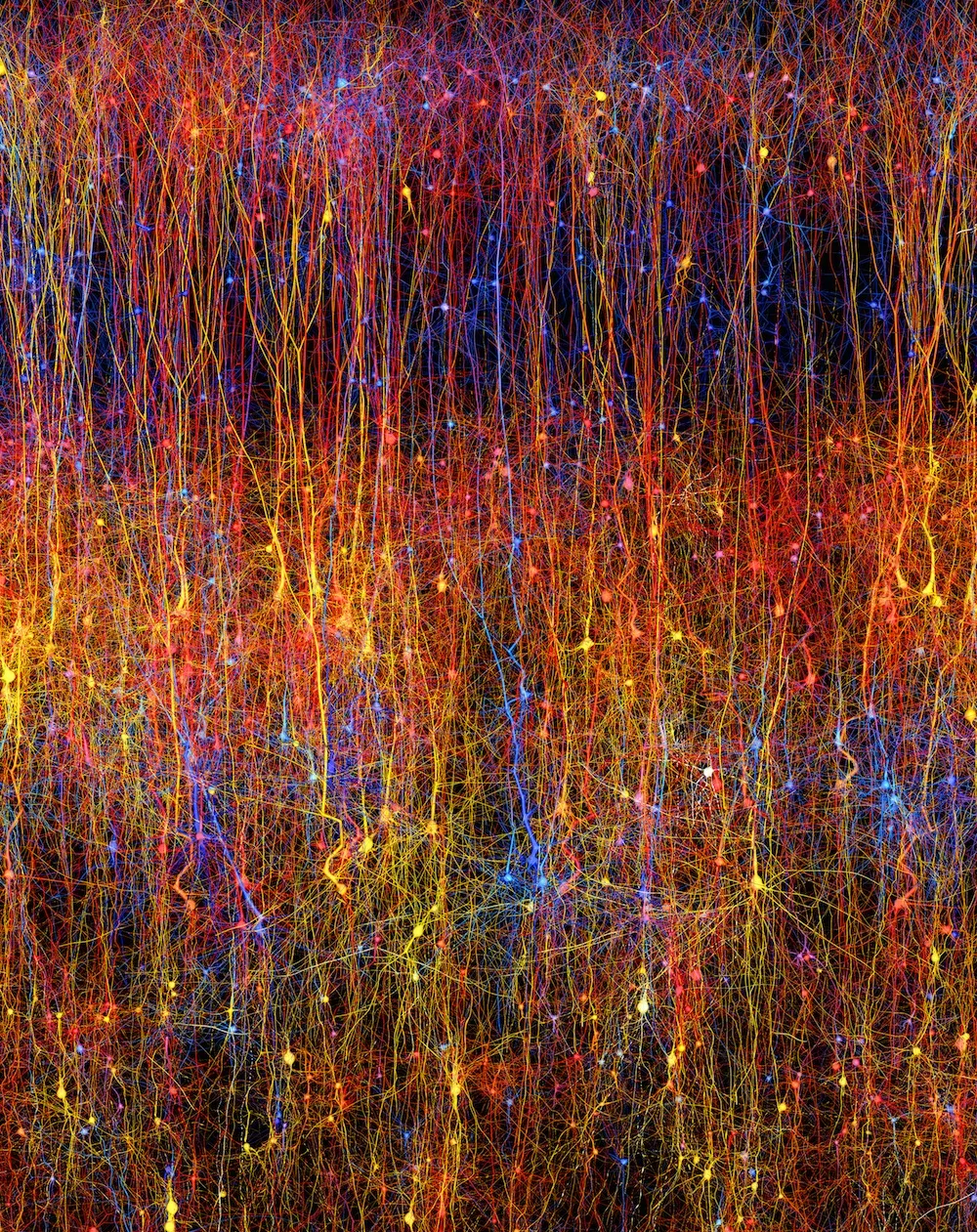
Here, another image of the digital rat brain slice, with red pyramidal cells, which play a key role in cognition, shown over a blue background network.
First draft
Still, the new model is only a first draft, and even for the tiny slice it captures just a fraction of the cells involved. For instance, the model doesn't represent support cells called glia, blood vessels, or the phenomenon of neuromodulation, in which brain chemicals can tune the behavior of many neurons. Here, another image of the digital rat brain.
Follow Tia Ghose on Twitter and Google+. Follow Live Science @livescience, Facebook & Google+. Original article on Live Science.

Tia is the editor-in-chief (premium) and was formerly managing editor and senior writer for Live Science. Her work has appeared in Scientific American, Wired.com, Science News and other outlets. She holds a master's degree in bioengineering from the University of Washington, a graduate certificate in science writing from UC Santa Cruz and a bachelor's degree in mechanical engineering from the University of Texas at Austin. Tia was part of a team at the Milwaukee Journal Sentinel that published the Empty Cradles series on preterm births, which won multiple awards, including the 2012 Casey Medal for Meritorious Journalism.