 Live Science Plus


Amazing fossil organs

Pumping blood
Fuxianhuia protensa, a 3-inch-long fossil found in sediments dating from 520 million years ago in China. This is an image of the animal's blood vessel system. The arthropod had a tubular heart located near the back of its body.
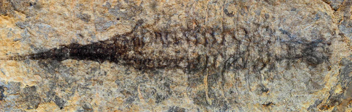
Hard shell

Arthropods are known for their protective exoskeletons — modern examples include lobsters and millipedes. This image shows the dorsal (back) view of Fuxianhuia protensa. Parts of the gut are visible as dark stains along the animal's midline.
Article continues belowCrustacean reconstruction

This image shows a schematic reconstruction of the shrimp-like fossil, outlining the cardiovascular system in red, the brain and central nervous system in blue and the gut in green.
Fossil head

The head region of Fuxianhuia from a fossil find reported in 2012, showing the brain and eye stalks.
Three-part brain

Iron-rich areas of the Fuxianhuia fossil, from 2012, reveal the imprint of an ancient brain.
Chengjiang arthropod

Fossil of the Alalcomenaeus, a megacheiran type of arthropod, a distant relative of scorpions and spiders.
Early brain

This close-up of the head region of the Alalcomenaeus fossil specimen includes superimposed colors of a microscopy technique that reveal the distribution of chemical elements in the fossil. Copper shows up as blue, iron as magenta and the CT scans as green. The researchers used CT scans to make 3D reconstructions of features of the fossilized nervous system. The scientists also used laser-scanning technology to map the distribution of chemical elements, such as iron and copper.

