
 Live Science Plus


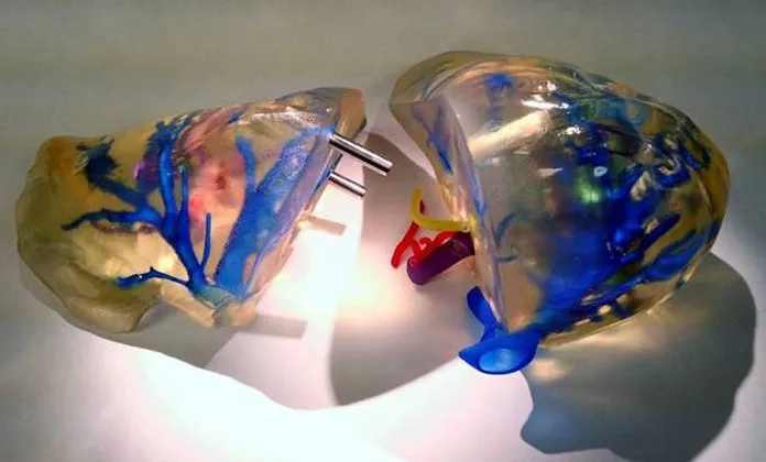
A new method of 3D printing an anatomically accurate replica of the human liver is now helping to guide surgeons during tricky procedures.
The 3D-printed replicas, which are made of transparent material threaded with colored arteries and veins, could help surgeons prevent complications when performing liver transplants or removing cancerous tumors, researchers said.
"We provide the surgeons with a physical model that is 100 percent identical to what they will encounter in surgery when they operate," said Dr. Nizar Zein, the chief of hepatology at the Cleveland Clinic in Ohio. "It takes away some of the potential surprises that will be found at the time of surgery."
The new liver replica could also be used to train medical students in the techniques needed for surgery, Zein said. [In Images of the 3D Printed Livers ]
Anatomically accurate
While reading a newspaper article about 3D printing, Zein realized that the technique could help make surgeries simpler. Before complicated liver surgeries, doctors usually look at a magnetic resonance image (MRI) or a computed tomography (CT) scan to visualize the liver and plan the operation.
But these 2D images don't provide true visual guidance during surgery. There are three main veins in the liver, and doctors often go into surgery unsure exactly where these blood vessels are located. Inadvertently cutting them can lead to "a disaster outcome," Zein told Live Science.
Get the world’s most fascinating discoveries delivered straight to your inbox.
To create the artificial livers, the researchers combine the MRI and CT scans that patients have already undergone, and then recreate the 3D shape of the organ. A study published last month in the journal Liver Transplantation confirmed that the models are anatomically accurate in terms of volume and location of vessels in the liver.
Using these models, the team creates the 3D-printed organs using a transparent polymer, then dyes the main blood vessels and the bile ducts.
Complications avoided
So far, the team has used such livers in about 30 cases. In a few operations so far, surgeons changed their plan for the surgery based on the simulated organs, for instance, after realizing that cancerous liver tumors were too close to certain veins to completely cut the growths out.
"We believe we actually avoided some complications this way," Zein said.
The researchers are now developing similar methods to guide complicated surgeries, such as hand and face transplants, and pancreatic tumor removals, Zein said.
They are also investigating a way to integrate organ models into the global positioning systems (GPS) that currently guide surgeries. These GPS tools determine the exact location to cut and the safe margins for a surgery. By improving the models of the organs that these systems use, the hope is that the GPS will become even more accurate, Zein said.
Follow Tia Ghose on Twitterand Google+. Follow Live Science @livescience, Facebook & Google+. Original article on Live Science.

Tia is the editor-in-chief (premium) and was formerly managing editor and senior writer for Live Science. Her work has appeared in Scientific American, Wired.com, Science News and other outlets. She holds a master's degree in bioengineering from the University of Washington, a graduate certificate in science writing from UC Santa Cruz and a bachelor's degree in mechanical engineering from the University of Texas at Austin. Tia was part of a team at the Milwaukee Journal Sentinel that published the Empty Cradles series on preterm births, which won multiple awards, including the 2012 Casey Medal for Meritorious Journalism.