Spotlighting the Ballet of Mitosis
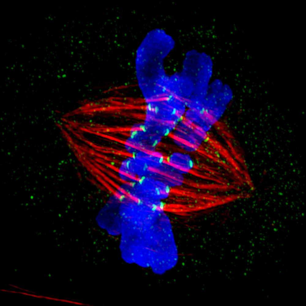
Like a pair of dancers sheathed in blue, two chromosomes take the cell's center stage in this scene from the elegant — and usually perfectly performed — process of mitosis.
Mitosis divides a single cell into two new cells, which is essential for cellular growth, reproduction and repair. During this delicate production, supporting dancers called spindle fibers, shown in red, ensnare the chromosomes, grasping them with the aid of harness-like structures called kinetochores, shown in green. Each chromosome is then gracefully escorted in opposing directions by the spindle fibers. This splits the duplicated genetic material in two for each of the new cells.
Indiana University researcher Jane Stout captured the stunning scene using a powerful OMX light microscope, which was funded through a National Institutes of Health grant to explore a number of important biological processes. Prior to the OMX microscope, scientists had the "cheap seats"— the best imaging tools could only depict the performers of mitosis as a bright, nebulous mass enclosed in a harried arrangement of overlapping lines.
The new microscope is like a ticket for orchestra seating. It uses a combination of four separate digital cameras and different colored lasers to take snapshots as frequently as every 10 milliseconds, producing three-dimensional images with extremely high resolution. These capabilities illuminate the locations of proteins involved in complex biological processes, including mitosis.
The increased detail will help scientists better understand what happens when the performance doesn't go as planned. Errors in mitosis can lead to unregulated cell division, as seen in many types of cancer.
The vivid images produced by the state-of-the-art system prompted the Indiana University scientists to dub it the "OMG microscope." Judges of the 2012 GE Healthcare Life Sciences Cell Imaging Competition were equally amazed. Stout's image was awarded first place in the high- and super-resolution microscopy category. As part of the prize, the image will be shown in high definition on an electronic billboard at 42nd Street and 7th Avenue in New York City's Times Square on Saturday, April 20, and on Sunday, April 21.
This Inside Life Science article was provided to LiveScience in cooperation with the National Institute of General Medical Sciences, part of the National Institutes of Health.
Get the world’s most fascinating discoveries delivered straight to your inbox.


