 Live Science Plus


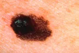
A new technique for imaging the deadly skin cancer melanoma could give surgeons clearer pictures of the cancer's boundaries, making it easier for them to ensure full removal of a tumor, researchers say.
Existing imaging techniques do not allow doctors to see melanoma's boundaries accurately enough to guide surgery, the researchers say, so surgeons tend to cut well beyond the visible tumor to be sure they remove all os the malignant tissue.
How it works
Bell discovered that the absorption of light causes material to heat up very slightly – perhaps only a few millionths of a degree. But this heat is enough to cause a material to expand.
"Much the same thing happens when you heat a balloon and it expands," study researcher Lihong Wang said in a statement.
If the light is pulsed at the right frequency, the material will expand and contract, generating a sound wave.
"We detect the sound signal outside the tissue," Wang said, and use computers to reconstruct the image.
Imaging techniques that rely on only on light waves have a limited ability to look deep into tissues, Wang said, because light is absorbed and scattered. But in tissue sound scatters much less than light, so photoacoustic tomography can "see" structures deeper inside the body.
The technique is safer than other means of deep imaging, because it uses rays that have lower energy levels than other imaging techniques, such as X-rays or positron emission tomography (PET) scans, according to the researchers.
Gold helps makes tumor boundaries clear
Photoacoustic images of melanoma tumors would have fuzzy edges without the use of a contrast agent. The researchers found the contrast is improved by loading the tumor with gold.
"Gold is much better at scattering and absorbing light than biological materials," said study researcher Younan Xia.
The gold nanoparticles used in the technique are bound to a hormone that binds to melanoma cells. Once injected, the gold particles naturally tend to accumulate in tumors, because the cells that line a tumor's blood vessels are disorganized and can leak contents.
Using the technique, the researchers reported that in mice they were able to clearly see melanomas below the skin that were barely visible to the unaided eye, and that the edges of tumors in the images were sharp.
It is estimated that 38,870 men and 29,260 women will be diagnosed with melanoma of the skin, and 8,700 people will die from it in 2010, according to the National Cancer Institute.
The work is published in the July issue of the journal ACS Nano.

