


Tiny Grandeur: Stunning Photos of the Very Small
Small Worlds

The Nikon International Small World Competition awards photographers for their renderings of the beauty and complexity of teensy things through the light microscope. The resulting photomicrographs must not only awe but also contribute significantly to various scientific disciplines. From alien-looking insects and worms to cells that would make stunning decorations, the 2011 award-winning photographs are both stunning and informative. Take a stroll into the world of the small.
Tiny Monster
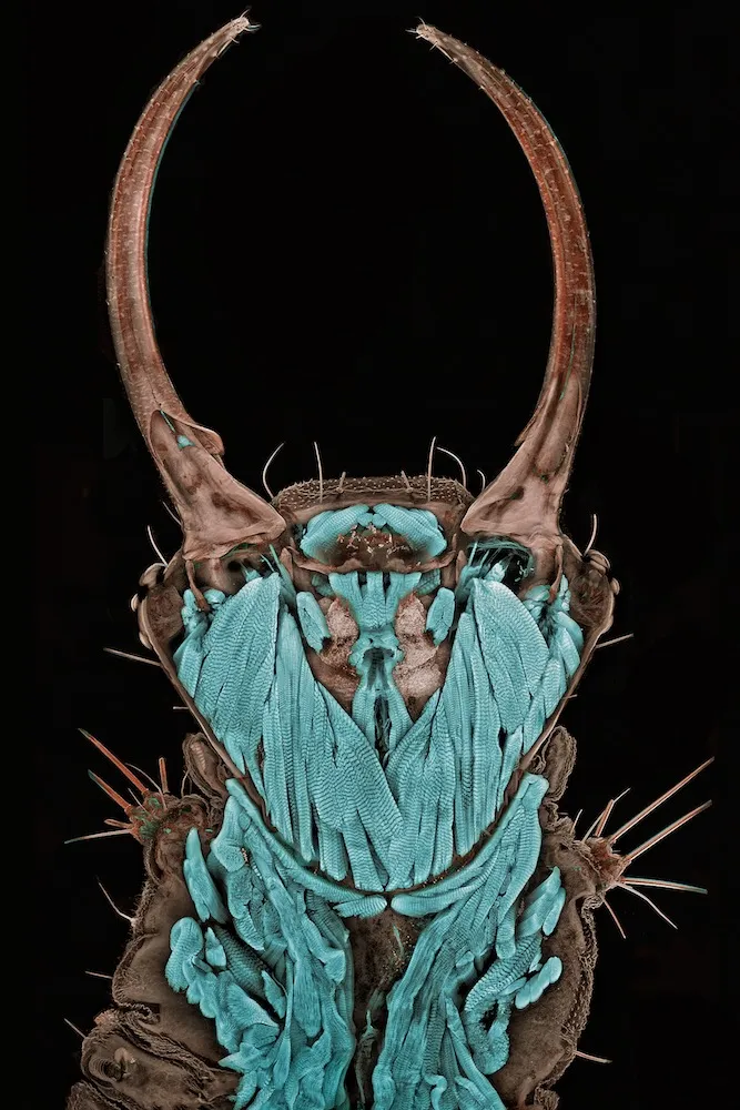
First place in the 2011 Nikon Small World photograph competition went to this photograph of an itsy-bitsy green lacewing larva. The bug landed on photographer Igor Siwanowicz and bit him; Siwanowicz retaliated by turning the insect into art.
Leaves of Grass

A blade of grass magnified 200 times took 2nd place in the Nikon Small World photography competition, 2011.

Bit of Algae

The third place winner of the 2011 Nikon Small World contest was this photo of living Melosira monliformis, a type of algae.
Fluorescent Beauty

Robin Young of the University of British Columbia took 4th place in the Nikon contest with this magnified image of liverwort.
A-maze-ing

Get ultra-close with electronics with this 5th-place winner of the Nikon Small World contest. This is the surface of a microchip.
Broken Windows

Sixth places in the Nikon Small World contest went to Dennis Callahan for this picture of cracked gallium arsenide solar cell films.
Get the world’s most fascinating discoveries delivered straight to your inbox.
Bright Lights

This image of mouse nerve fibers lit up the judges and won 7th place in the 2011 Nikon Small World contest.
Stained Glass Rocks

Italian geoscientist Bernardo Cesare took 8th place in the Nikon contest for this close-up image of a graphite-bearing granulite from India. Cesare talked to LiveScience about his work in August; see more of his beautiful photography here.
Itsy-Bitsy Ocean-Dweller

The marine copepod Temora longicornis takes on subtle hues in this 9th-place-winning photograph.
Hello, Flea

Don't be bashful, little flea -- you've won 10th place in the 2011 Nikon Small World contest. This is a freshwater water flea, Daphnia magna.
