 Live Science Plus


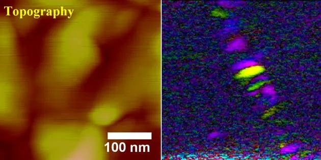
Scientists have successfully imaged tiny biological structures that are normally hidden by surrounding material.
The structures are less than 150 nanometers across. The details in these images can be less than 10 nanometers. That's 10 billionth of a meter, just the width of a handful of atoms laid end-to-end.
The structure in question was a single protein fiber that was embedded in tooth enamel. But this technique could work with any human, animal, or plant tissues, says Sergei Kalinin, a research scientist at the Oak Ridge National Laboratories in Oak Ridge, Tennessee.
Kalinin and his colleagues at North Carolina State University in Raleigh form the images by harnessing the piezoelectric effect. Piezoelectric materials either move when an electric current is applied to them or produce an electric current when they are compressed. Perhaps the best known piezoelectric materials are quartz crystals, whose electricity-prodded vibrations controlled oscillators in watches and early radios.
Many biological materials, such as bones, tendons, and wood, also move slightly when electrically shocked.
Using a custom-built tip extension for a scanning force microscope, the scientists direct a tiny voltage, which alternates polarity 50,000 times per second, at small groups of piezoelectric-sensitive molecules. The molecules then vibrate 50,000 times per second while the surrounding non-piezoelectric materials remain still.
By tracking patterns the vibrating molecules make, the scientists produce images of tiny structures that otherwise would be lost among other, non-piezoelectric, materials, such as hydroxyapatite, which is a type of calcium.
This technology, Kalinin says, works at a material's surface. Although the most likely near-future applications are in fundamental research, he says, it is possible that someday it will allow faster and cheaper analysis of biopsy samples. Current imaging technologies require technicians to spend time staining biopsy samples. The new technique wouldn't require a stain.
Another possible future application would be to image and then use the same tool, at a higher voltage, to selectively zap viral contaminants from biological samples.
"One of the things we have done recently is to use electrical bias to selectively modify, for example, the tobacco mosaic virus," Kalinin explained. The virus affects flowers and vegetables worldwide.
"If we have viruses on the surface, we can see them," he said. "Secondly, we can select the viruses we don't like and blow them up by applying a high enough electric field."
- Gallery: Micromachines
- World's Smallest Hypodermic Needle
- Gold Probes Could Reveal Cancer in Your Body
- Microscopic Images as Art
