It's quick and easy to access Live Science Plus, simply enter your email below. We'll send you a confirmation and sign you up for our daily newsletter, keeping you up to date with the latest science news.
A team of neuroscientists has produced a series of amazing, detailed images of fruit fly brains.
The images aren't quite photos, but they were made by capturing visible light. To create them, the researchers combined two techniques — one that caused the brain tissue to grow much larger than its usual size, and another that allowed the researchers to make precise photos of that tissue without damaging it. [Magnificent Microphotography: 50 Tiny Wonders]
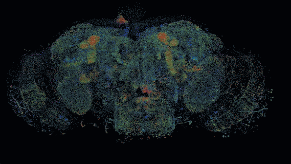
This is a map of dopaminergic neurons in the right hemisphere of a fruit fly brain.
(Image credit: Courtesy of the researchers)
The result was a colorful and fully searchable map of a fruit fly brain, which according to a statement from MIT (where one of the researchers works) is no bigger than a poppy seed.
Making delicate tissues expand is a tricky business, but it can be useful for neuroscience research; in many circumstances neurons and their connections are too tiny to easily image and map. The technique, called "expansion microscopy," first emerged in 2015, detailed in a paper by Ed Boyden (one of the creators of the fruit fly images and a neuroscientist at MIT) and two other researchers.
To make the technique work, they found a polymer that would enter cells without destroying them. Then, they soaked a mouse brain in the stuff. Once the polymers permeated the tissue, the researchers poured a bath over the tissue that caused the polymers to expand, physically expanding the cells themselves for easier study.
Another image shows neurons in the fruit fly brain, color coded by depth.
(Image credit: Courtesy of the researchers)
That technique alone wouldn't have been enough, however, to create these beautiful brain images. To scan the expanded brain in enough detail, the researchers used a technique previously developed by another co-author — Eric Betzig, a biologist at UC Berkeley — for rapidly 3D-scanning tissues using only light and microscopes.
That technique, called "lattice light sheet microscopy," involves shining a line of light up through the bottom of the tissue. It lights up just one flat plane of the tissue, as if a single slice started glowing within a loaf of bread, bright enough to be seen through the front of the loaf. A microscope camera mounted at a 90-degree angle to the beam of light is then able to spot that illuminated plane and record what it looks like. Do that over and over again (from the front slice to the rear), and you're left with a three-dimensional image of the tissue.
Sign up for the Live Science daily newsletter now
Get the world’s most fascinating discoveries delivered straight to your inbox.
This image, taken from a mouse brain, shows neurons (yellow) and proteins involved in their synapses (cyan and magenta).
(Image credit: Courtesy of the researchers)
This is a big deal, the researchers said, because both expansion microscopy and lattice light sheet microscopy are relatively quick and straightforward methods for neuroscientists to use in their labs. And now, combined, they can allow researchers to rapidly image large chunks of brain in incredible detail.
Neuroscience is increasingly concerned with understanding large portions of the brain without letting go of a microscope-level view of what's going on. Some researchers think that mapping the brain in detail could unlock its secrets. Now, they have a new way to do that.
Rafi joined Live Science in 2017. He has a bachelor's degree in journalism from Northwestern University’s Medill School of journalism. You can find his past science reporting at Inverse, Business Insider and Popular Science, and his past photojournalism on the Flash90 wire service and in the pages of The Courier Post of southern New Jersey.