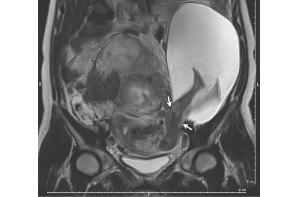
Just looking at this image might give the impression that this woman's baby literally kicked its feet right out of her uterus. But moms-to-be with kicky babies can rest easy — the MRI image showcases an extremely rare condition that was not caused by a baby's kick.
The 33-year-old woman had developed a 1 inch (2.5 centimeters) tear in the wall of her uterus, and through the tear, part of the amniotic sac measuring 7.5 by 4.7 by 3.5 inches (19 by 12 by 9 cm) popped out, according to a brief report of her case. The amniotic sac is the fluid-filled membrane found in the uterus that contains the growing and developing fetus.
But the woman had no symptoms that any of this was going on. She didn't learn of her condition until she came in for a routine ultrasound when she was 22-weeks pregnant, according to the report, published today (Dec. 21) in The New England Journal of Medicine.
Latest Videos From
Dr. Pierre-Emmanuel Bouet, an OB/GYN at the Angers University Hospital in France and the lead author of the report, said he had never seen a case like this before. [Here's a Giant List of the Strangest Medical Cases We've Covered]
Indeed, the condition is "extremely rare," Bouet told Live Science. There have only been 26 cases reported in the literature, he added.
This was the woman's sixth pregnancy, the doctors wrote in the report. In all of her five previous pregnancies, the woman delivered the babies via Caesarean section (C-section), they wrote.
In fact, it was the woman's five previous C-sections that increased her risk for a uterine tear, Bouet said. It seems that her C-sections had weakened the wall of the uterus, he said. The tear didn't occur at the exact location of the earlier C-sections, but close by, he added.
The area of the uterus that had scarred after the C-sections was strong, but the regions around this scar were fragile, Bouet said. The forces and pressures on the uterus that occur during pregnancy ultimately led to the tear, he said.
Upon discovering the woman's uterine tear and protruding amniotic sac, the doctors informed the woman and her husband of the potential risks, which included additional uterine tearing, preterm birth and a serious pregnancy complication called placenta accreta, in which the placenta doesn't detach from the uterine wall after birth.
It was also possible for the amniotic sac to rupture, Bouet said. If this occurred, the doctors would make sure that the fetus still had a heartbeat, and if so, would perform an emergency C-section, Bouet said. The doctors would also have to consider the age of the fetus: if it was too early in the pregnancy, the odds of survival would be lower, he said.
The woman and her husband decided to continue the pregnancy with close monitoring, according to the report. Bouet said that the woman was not put on bed rest during this time, and that she could do some moderate walking.
By 30 weeks, the tear in the woman's uterus grown by 2 inches (5 cm) and the portion of the amniotic sac outside the uterus had grown in size, the doctors wrote in the report. At that point, not only did this part of the amniotic sac contain the fetus's legs, but also the abdomen, they wrote.
The doctors and the woman decided to deliver the baby via C-section. The baby boy was healthy, and weighed in at 3 lbs. (1.385 kilograms), according to the report. After delivery, the doctors repaired the woman's uterus, and she returned home from the hospital after five days.
The doctors last checked in with mother and baby six months after he was born, and noted that they were doing well.
Originally published on Live Science.

 Live Science Plus