Ancient Spider Guts Revealed in 3-D
Get the world’s most fascinating discoveries delivered straight to your inbox.
You are now subscribed
Your newsletter sign-up was successful
Want to add more newsletters?
Delivered Daily
Daily Newsletter
Sign up for the latest discoveries, groundbreaking research and fascinating breakthroughs that impact you and the wider world direct to your inbox.
Once a week
Life's Little Mysteries
Feed your curiosity with an exclusive mystery every week, solved with science and delivered direct to your inbox before it's seen anywhere else.
Once a week
How It Works
Sign up to our free science & technology newsletter for your weekly fix of fascinating articles, quick quizzes, amazing images, and more
Delivered daily
Space.com Newsletter
Breaking space news, the latest updates on rocket launches, skywatching events and more!
Once a month
Watch This Space
Sign up to our monthly entertainment newsletter to keep up with all our coverage of the latest sci-fi and space movies, tv shows, games and books.
Once a week
Night Sky This Week
Discover this week's must-see night sky events, moon phases, and stunning astrophotos. Sign up for our skywatching newsletter and explore the universe with us!
Join the club
Get full access to premium articles, exclusive features and a growing list of member rewards.
Digital wizardry has allowed scientists to see the insides of a 53-million-year-old fossilized spider in 3-D.
The male spider is about the size of a pinhead (or a stack of three salt grains) and lived during the early Eocene epoch, from about 55 million to nearly 34 million years ago.
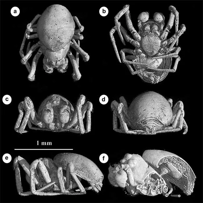
It represents a new genus and species dubbed Cenotextricella simoni. It's also the earliest fossil species of a family of micro-spiders called Micropholcommatids from Australia, New Guinea, New Caledonia, New Zealand and Chile.
The fossil was found preserved in amber in the Paris Basin in France.
“Amber provides a unique window into past forest ecosystems," said lead author David Penney, a paleo-arachnologist at the University of Manchester in England. "It retains an incredible amount of information, not just about the spiders themselves, but also about the environment in which they lived."
Normally, observing the minute features of an already tiny specimen would mean cracking into the amber, potentially destroying the sample.
Instead of physical dissection, Penney and his colleagues used Very High Resolution X-ray Computed Tomography to scan the bug through its amber grave.
Get the world’s most fascinating discoveries delivered straight to your inbox.
The resulting 3-D reconstructions could be sectioned and viewed from various angles, essentially allowing for digital dissection of the spider specimen.
“This technique essentially generates full 3-D reconstructions of minute fossils and permits digital dissection of the specimen to reveal the preservation of internal organs," Penney said.
- Video: Mating Spiders
- Vote: The Ugliest Animals
- Image Gallery: Creepy Spiders
Jeanna Bryner is managing editor of Scientific American. Previously she was editor in chief of Live Science and, prior to that, an editor at Scholastic's Science World magazine. Bryner has an English degree from Salisbury University, a master's degree in biogeochemistry and environmental sciences from the University of Maryland and a graduate science journalism degree from New York University. She has worked as a biologist in Florida, where she monitored wetlands and did field surveys for endangered species, including the gorgeous Florida Scrub Jay. She also received an ocean sciences journalism fellowship from the Woods Hole Oceanographic Institution. She is a firm believer that science is for everyone and that just about everything can be viewed through the lens of science.
