


Gallery: Trippy Photos Reveal Beauty in Science
Natural Beauty

Here's a look at some of the winners of the 2012 Science and Engineering Visualization Challenge, whose photos, interactive videos and even computer games reveal the beauty of the natural world.
Cognitive Connectivity

Cognitive Computing researchers at IBM are developing a new generation of "neuro-synaptic" computer chips inspired by the organization and function of the brain. For guidance into how to connect many such chips in a large brain-like network, they turn to a "wiring diagram" of the monkey brain as represented by the CoCoMac database. In a simulation designed to test techniques for constructing such networks, a model was created comprising 4173 neuro-synaptic "cores" representing the 77 largest regions in the macaque brain. The 320749 connections between the regions were assigned based on the CoCoMac wiring diagram. This visualization is of the resulting core-to-core connectivity graph. Each core is represented as an individual point along the ring; their arrangement into local clusters reflects their assignment to the 77 regions. Arcs are drawn from a source core to a destination core with an edge color defined by the color assigned to the source core.
Cerebral Infiltration

The image is the result of fiber tractography from diffusion-weighted magnetic resonance imaging. It illustrates the white matter of the brain, or in other words, its structural connections. The red smooth surface represents a glioblastoma tumor. We can see the effect of repulsion and infiltration of this mass on the white matter fiber pathways. A distance colormap is used for interpretation. Blue fibers mean that they are located within a safe distance of the tumor whereas red fibers are in a close perimeter to the tumor, and can cause severe post-operation deficits, if resected.

Plant Seeds

High-resolution high-contrast X-ray radiography of plant seeds combined with images taken by microscopy. The X-ray images were measured using combination of a micro-focus X-ray source and a state-of-the-art hybrid pixel semiconductor detector. The detector enables imaging in so-called single photon counting regime allowing acquiring radiographs with theoretically unlimited dynamic range (in practice limited just by the number of detected photons). In combination with point-like source magnifying geometry, the technique presents a powerful tool allowing nondestructive investigation of mm-sized object of any kind. The results show a novel application of the technique to plant biology, namely the visualization of seeds (typically 3 mm in size). For better interpretation of imaged features, the radiographs are combined with the images taken by microscopy.
Biomineral Single Crystals

Biomineral crystals found in a sea urchin tooth. Geologic or synthetic mineral crystals usually have flat faces and sharp edges, whereas biomineral crystals can have strikingly uncommon forms that have evolved to enhance function. The image here was captured using environmental scanning electron microscopy and false-colored. Each color highlights a continuous singlecrystal of calcite (CaCO3) made by the sea urchin Arbacia punctulata, at the forming end of one of its teeth. Together, these biomineral crystals fill space, harden the tooth, and toughen it enough to grind rock.
Self Defense
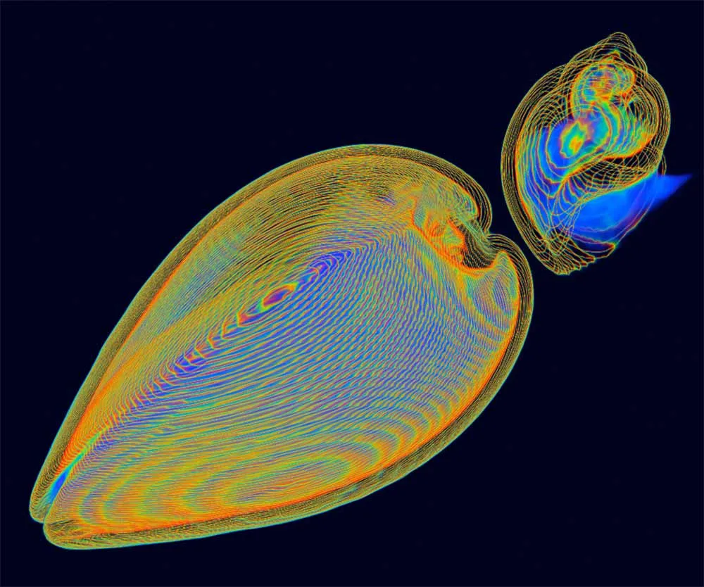
Evolution encourages diversity, allowing Nature to solve problems in more than one way. Thisimage is a 3D CT scan of a clam and a whelk, both alive. The clam (left) is nestled comfortablyin the bottom half of its shell. Note the simplicity of the hinge design in its bivalve shell. Byclosing the shell rapidly, the clam is able to fence off a potential attack. Yet the whelk's shell(right) is even more amazing. The sophisticated spiral construction is astonishingly complex andstrong, an architectural marvel by itself and an evolutionary success! Once the whelk slippedback into the spiral tunnel of its shell, the shell provides protection similar to a fortress. Both theclam and the whelk solve the vital problem of self defense, albeit in different ways. The whelkhowever has the upper hand because it has the ability to drill a hole directly through the clam'sshell by softening it with secretions and then consumes the clam as meal.
A Computational Heart

Here, a screenshot of a video on the complex and fascinating organ – the heart. Scientists hope to simulate the beating heart realistically and in the video they describe a project called Alya Red, aimed at developing a computational cardiac model. The tone of the video is educational, although the renderings are of actual simulation results.
Get the world’s most fascinating discoveries delivered straight to your inbox.
Owl Rotation

Owls (Order Strigiformes) can perform 270-degree neck rotations. The cervico-cephalic vesselsare notoriously sensitive to rotary motion in most vertebrates, including man, in whom injury ofthese arteries commonly leads to cerebral infarction. This poster was created as part of aMaster’s thesis study that examined whether owls have evolved specific arterial adaptations thataccommodate their extreme range of neck rotation. The intermediate carotid and vertebralarteries were closely examined from the basi-cervical region up to the formation of the basilarartery using 3D Fusion digital subtraction angiography and traditional dissection techniques.Numerous vascular adaptations were documented that were considered directly related to neckrotation. The study was conducted on 12 deceased owl specimens. None were sacrificed for thepurpose of this study. The full study team included Fabian de Kok-Mercado, Michael Habib,Tim Phelps, Lydia Gregg and Philippe Gailloud.
Earth Evolution

This educational poster shows how geological and biological processes have together shaped Earth’s environment during its 4.6 billion-year history.
In a Mouse's Eye
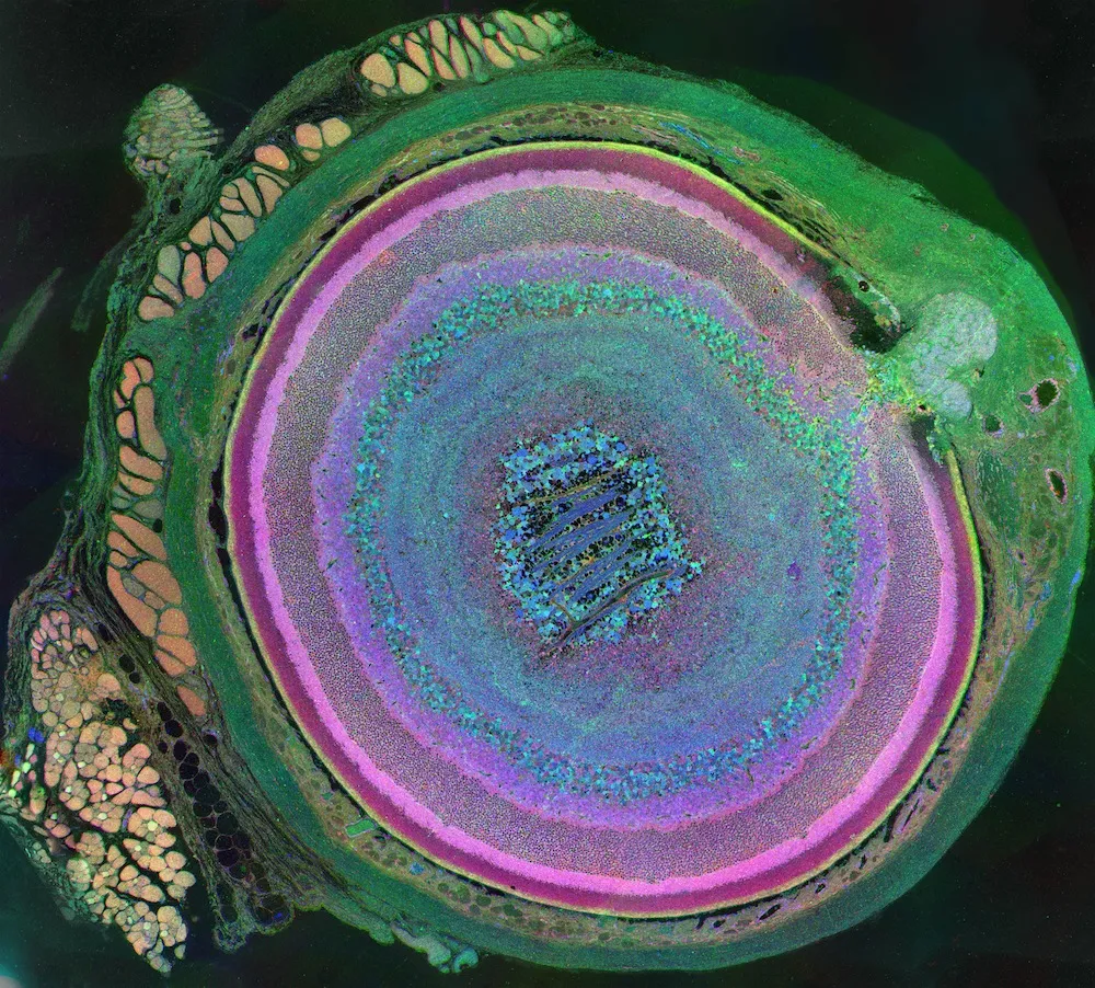
Here, a winner in last year's challenge. This computational molecular phenotype image of a mouse's eye reveals the diversity of cell metabolism in the retina. The optic nerve is in the upper right of the image. The rectus muscles can be seen in red and gold, attached to the green sclera (the white part of the eye). Retinal layers appear in a rainbow of colors from light gold to pink and purple, while other cells show up in blue and green.
Cool as a ...

Another 2011 winner: This 2011 honorable mention photo is the skin of an immature cucumber, magnified 800 times. These structures are called "trichomes," and they act as little spears, protecting the young vegetable from plant-eaters. The lower part of the trichomes contains bitter, toxic chemicals that make herbivores go "ick!" [See more images from last year's winners]
