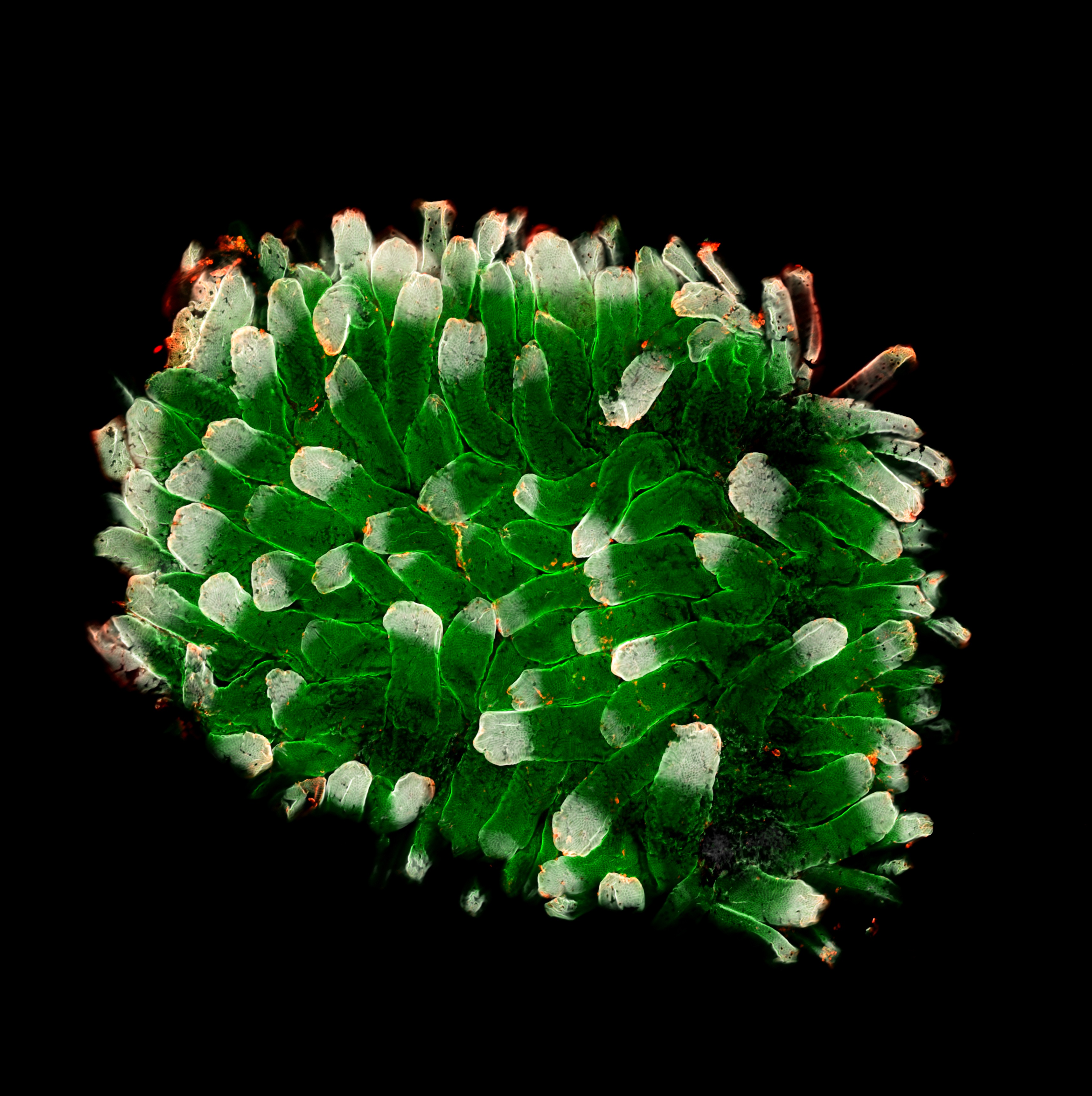
This mysterious green spongelike structure is actually an up-close look at a section of a mouse's small intestine.
Bryan A. Millis captured this cool microphotograph, which was awarded "Honorable Mention" at 2013 Nikon Small World Competition. Millis stitched this large format image using swept-field confocal fluorescence microscopy at 200 magnification.
Autofluorescence is the natural emission of light by biological structures when exposed to light they absorb, in this case the small intestinal section from a mouse expressing GFP-tagged non-muscle myosin II. Most biological structures and even some synthetic products, such as paper, have some degree of autofluorescence. Autofluorescence from U.S. paper currency is used to discern counterfeit from authentic money.
Millis also used confocal optical imaging to improve detail in the image. The technique aims at eliminating out-of-focus signals from the microscope. A conventional microscope "sees" as far into the specimen as the light can penetrate, while a confocal microscope only "sees" images one depth level at a time making each more controlled and focused. By stacking several images, Millis is able to get a close-up look at the intestine.
Millis is a cell biologist at the National Institute on Deafness and Other Communication Disorders, Laboratory of Cell Structure and Dynamics, National Institutes of Health (NIH) in Bethesda, Md.
Follow Live Science @livescience, Facebook & Google+.
Get the world’s most fascinating discoveries delivered straight to your inbox.
 Live Science Plus


