Image Gallery: The Oddities of Human Anatomy
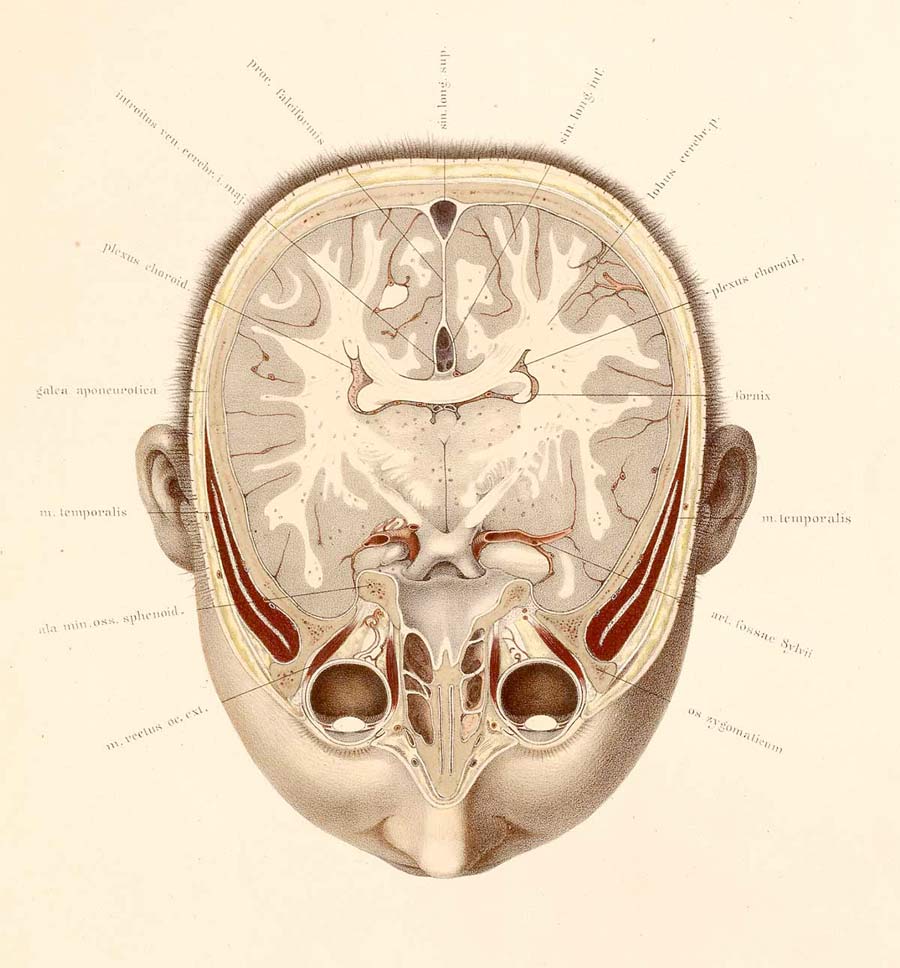
A new science of human anatomy arose some 500 years ago, with imagery that was both informative and whimsical, surreal, beautiful and grotesque, according to the National Library of Medicine, whose exhibition "Dream Anatomy" reveals the amazing anatomical imagery.
Here, a cross-section from the atlas of anatomist Wilhelm Braune and artist C. Schmiedel.
The female body
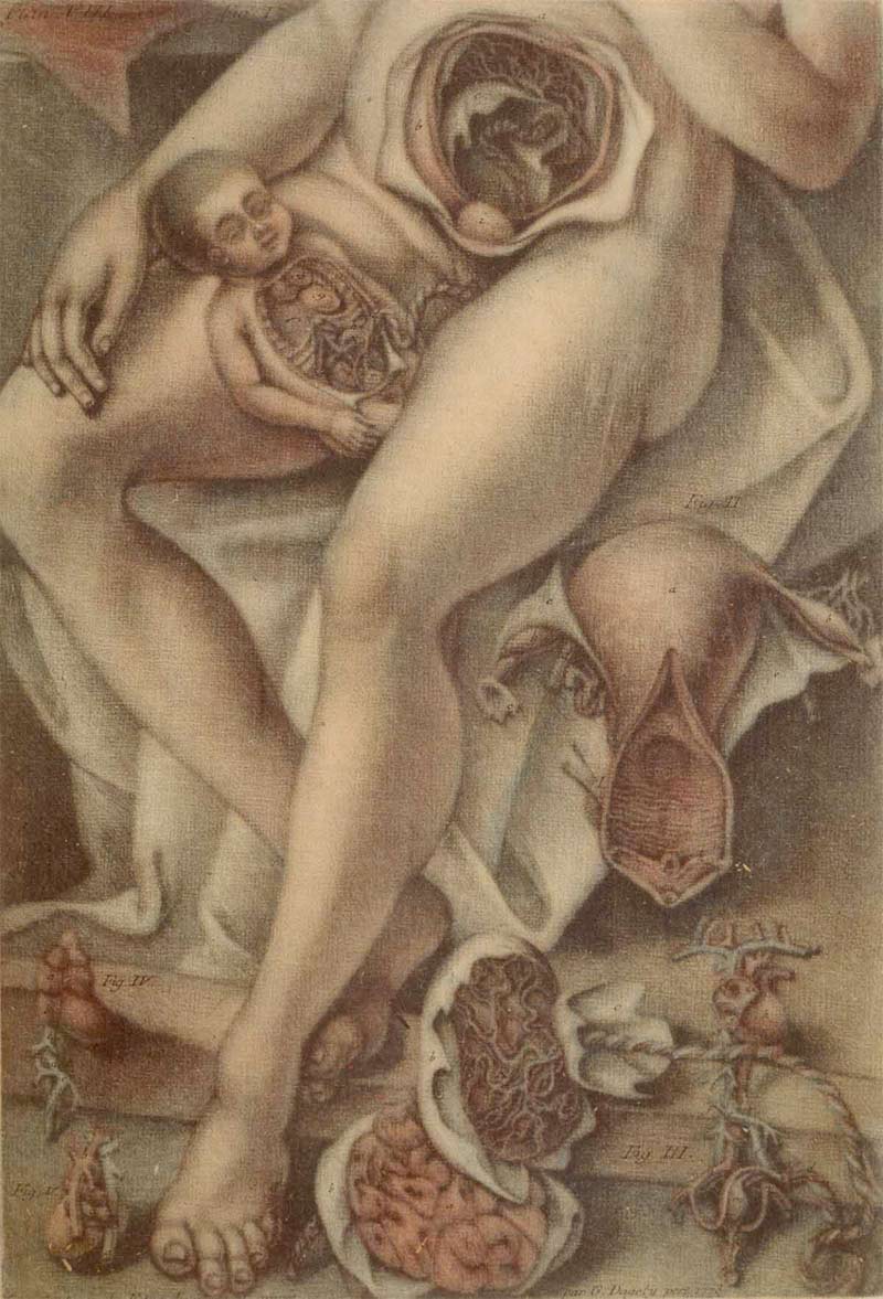
This colored mezzotint by author and artist Jacques Fabien Gautier D'Agoty reveals "the grotesquerie of subject matter, stiffness of the figure, and eccentric arrangement of body parts make for a characteristic dreaminess that eerily anticipates 20th-century modernism," the National Library of Medicine states.
Life and Death
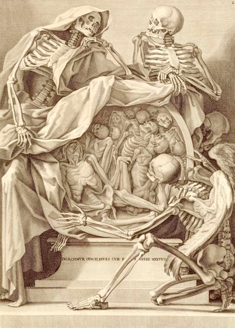
The association between death and anatomy continued in art anatomy, even as it waned in medical texts, as shown here. Bernardino Genga, a Roman anatomist, specialized in studies of classical sculptures, while Charles Errard, court painter to Louis XIV, helped found the Académie Royale de Peinture and was first Director of the Académie de France in Rome.
Anatomical Manakin
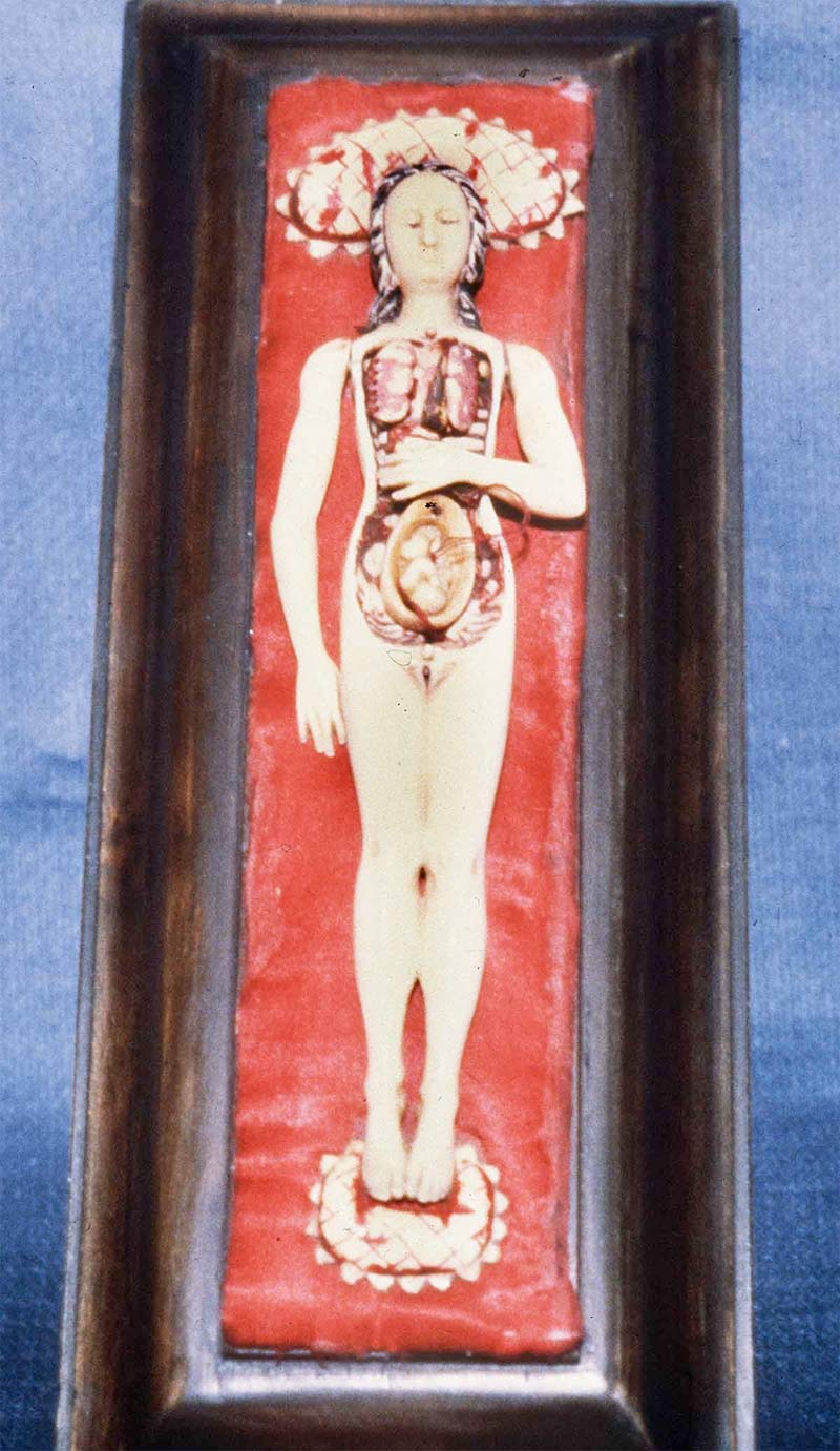
These manikins, between 6 to 7 inches in length, were made from solid pieces of ivory some time between 1500 and 1700. The arms were carved separately and are moveable. The thoracic and abdominal walls can be removed, revealing the viscera. In some manikins the internal organs are carved in the original block and are not removable, while they are formed into separate pieces that can be removed.
Facial Arteries
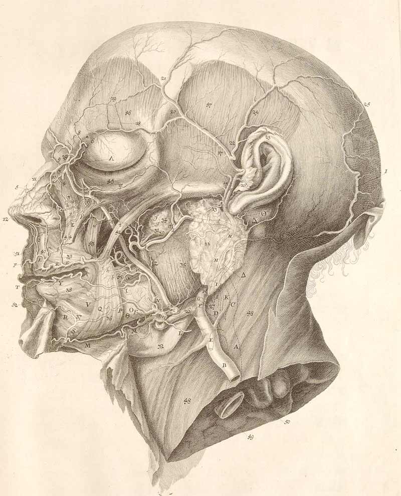
Contemporaries praised the Swiss anatomist Albrecht von Haller for his finely detailed illustrations of finely dissected subjects. This dissection of the arteries of the face was copied and reprinted in numerous other works of anatomy. (Artist: C.J. Rollinus)
Dancing Skeleton
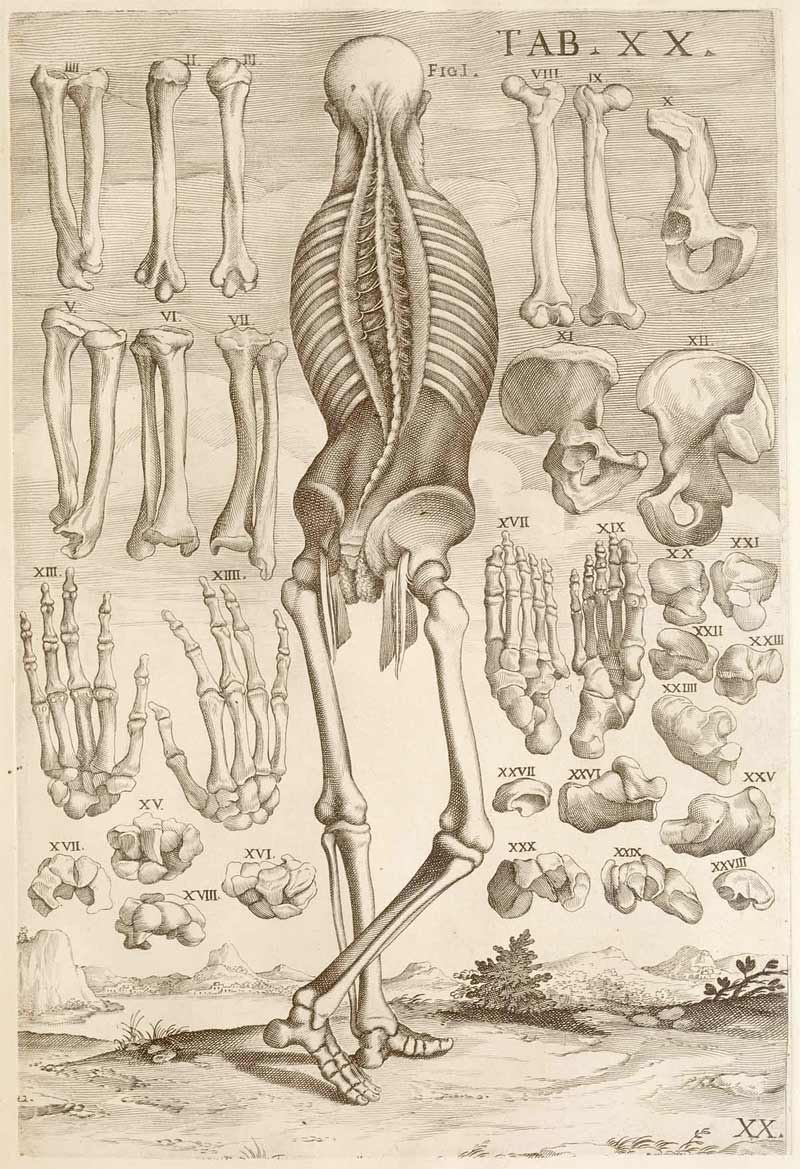
"A skeleton dances a lively step; in the background an arrangement of bones float in the air," according to the National Library of Medicine. Pietro Berrettini da Cortona's "exuberant flourishes take their cue from the theatricalism of baroque drama and court entertainments."
Disembodied Legs
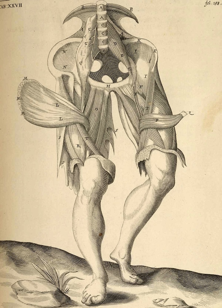
The muscles of the thigh are illustrated in this drawing reminiscent of men's breeches.
Get the world’s most fascinating discoveries delivered straight to your inbox.
A New World
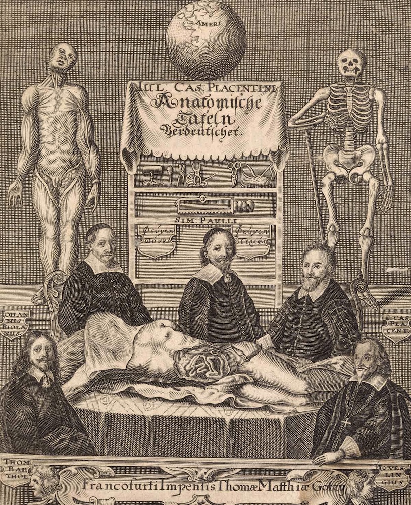
A frontispiece portrays five anatomists posed around a cadaver. The globe at the top of the illustration, turned toward America, reveals how the anatomists saw themselves: as exploring a "New World" of science.
Colorful Muscles
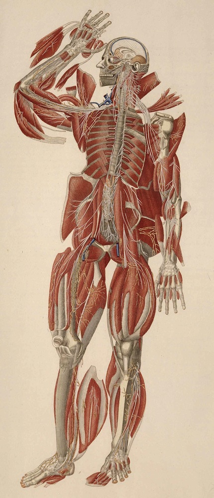
Colorful images came into fashion in the 1800s, but the flap-like dissection of the muscles heralds back to older styles of anatomical art.
Harsh Realities
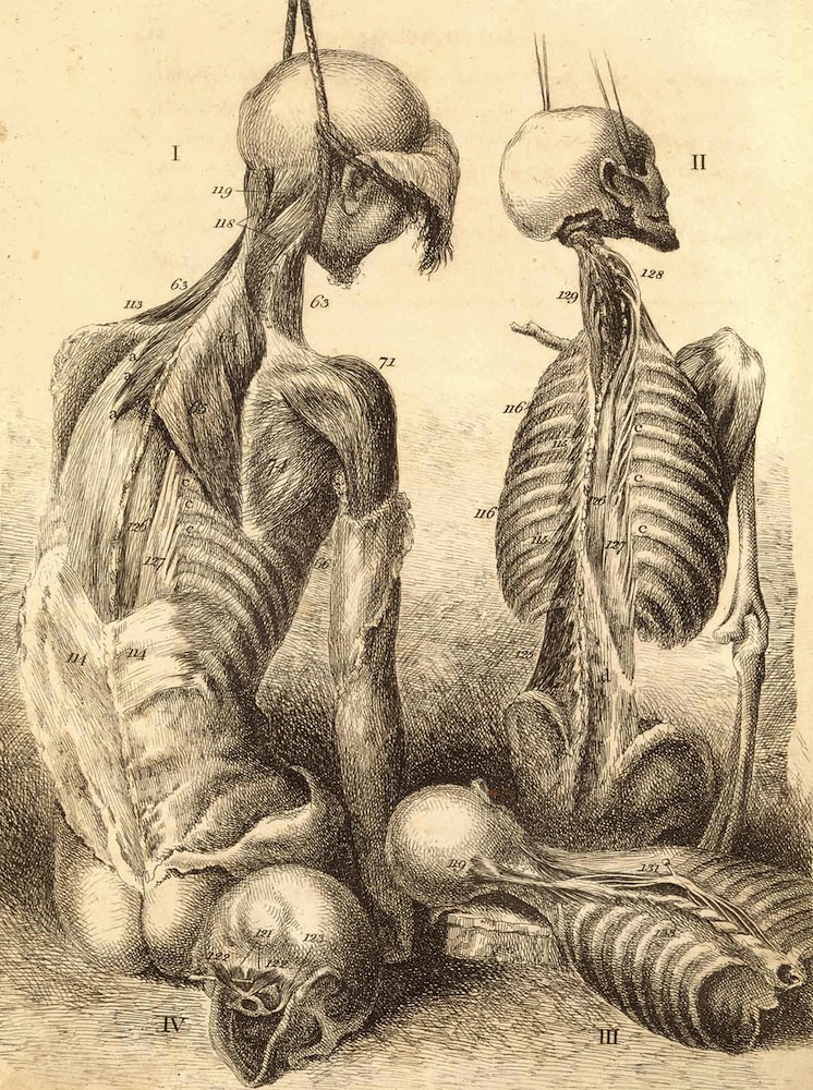
Artist John Bell decried overly idealized anatomical art, preferring the harsh realities of dissection.
Pregnancy Pose
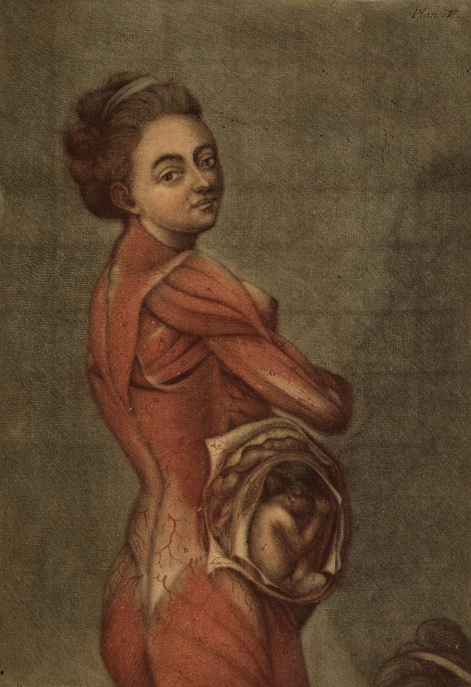
A classic pose of French portraiture meets anatomical art in this painting of a pregnant woman from 1773.
 Live Science Plus


