


Why Skin Is Strong: Cells Stick Like Velcro
Scientists have gotten their best look ever at interactions inside human skin cells, finding a Velcro-like setup that links them and makes skin strong while also supple.
The cell-interior images, made with a new a technique called cryo-electron tomography, show the proteins responsible for cell-cell contacts for the first time.
"This is a real breakthrough in two respects," said Achilleas Frangakis of the European Molecular Biology Laboratory. "Never before has it been possible to look in three dimensions at a tissue so close to its native state at such a high resolution. We can now see details at the scale of a few millionths of a millimeter. In this way we have gained a new view on the interactions of molecules that underlie cell adhesion in tissues—a mechanism that has been disputed over decades."
The results are detailed in the Dec. 6 issue of the journal Nature.
So far, the only information available about a protein’s position and interactions in a cell was based on either light microscopy images at poor resolution or techniques that remove proteins from their natural context. Electron microscopy normally requires tissue to be treated with chemicals or coated in metal, a procedure that disturbs the natural state of a sample.
Frangakis and his group developed a technique that instantly freezes cells in their natural state prior to imaging with an electron microscope. With cyro-electron tomography, images of the untreated sample are taken from different directions and assembled into an accurate 3-D image by a computer.
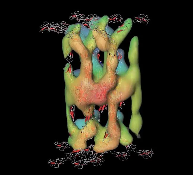
The researchers applied this technique to observe proteins that are crucial for the integrity of tissues and organs such as the skin and heart, but also play an important role in cell proliferation. These proteins, called cadherins, are anchored in cell membranes and interact with each other to bring cells close together and interlink them tightly.
Get the world’s most fascinating discoveries delivered straight to your inbox.
"We could see the interaction between two cadherins directly, and this revealed where the strength of human skin comes from," says Ashraf Al-Amoudi, who carried out the work in Frangakis’ lab. "The trick is that each cadherin binds twice: once to a molecule from the juxtaposed cell and once to its next-door neighbor. The system works a bit like specialized Velcro and establishes very tight contacts between cells."
- Image Gallery: Microscopic Images as Art
- DNA Art: Origami Goes Nano
- Living Cells Scanned in 3-D for First Time
