


Science as Art: A Gallery
Meteorite Pop Art

Scientists use X-rays, dyes, fancy microscopes and other tools to see things we can't capture with our naked eyes. But these tools aren't just good for science, they can make art. An exhibit at the American Museum of Natural History explores the beauty in scientific imaging. These pictures illustrate the chemical composition of four meteorites, which was detected by scanning them with a beam of electrons. Red represents magnesium, green is calcium, and blue is aluminum.
Hidden Contents

Museum conservators use X-rays to investigate the condition of artifacts, including this wooden Tibetan deity figure. The scan revealed previous repairs as well as ritualistic objects placed within a cavity inside the body.
No Dissection Needed

The bones and cartilage in this Madagascar cichlid fish, which was still whole when the image was made, stand out sharply thanks to red and blue dye, as well as other chemicals that rendered the rest of the fish's body transparent.

Spook Alley Scorpions

Under ultraviolet light, these scorpions glow naturally. A museum arachnologist takes advantage of this trait to identify differences among the species shown.
Up Close And Personal

Close-ups of bugs' private parts, like the male genitalia of a Peruvian shore bug, help scientists identify the species to which they belong. This image was made with a scanning electron microscope, which sweeps a beam of electrons (negatively charged particles in atoms) across the sample’s surface.
Fluorescent Coral

This staghorn coral contains fluorescent molecules that absorb light from an external source — in this case, the sun — and then emit light at a different wavelength. Museum zoologist David Gruber takes advantage of this to take close-up images of corals. These form part of his research into the patterns and function of fluorescent proteins. Some of these proteins have been co-opted as important tools in biomedical research.
Bacteria in Leeches

Scientists used special DNA to label the bacterial symbionts living within leeches in order to see them under a microscope.
Get the world’s most fascinating discoveries delivered straight to your inbox.
Armadillo Lizard
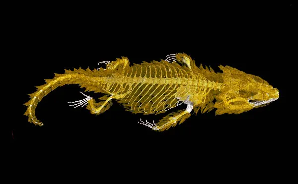
This portrait of an armadillo lizard, captured by the X-rays of a CT scanner, shows the animal's bony plates protecting its body.
Volcanic Cross Section

This cross section of two volcanic craters on Jan Mayen Island in the North Atlantic Ocean was revealed when a massive amount of rock fell into the ocean. The reddish color, intensified in the photo through image processing, indicates age-old deposits of volcanic rocks containing oxidized iron.
Discovering a Star

Astrophysicists discovered a star in the Big Dipper with the help of advances in a technique called coronagraphy. With it, bright light from a star can be blocked out better than ever before, so that faint nearby objects can be seen. As a result, the scientists determined that a faint red dwarf, now known as Alcor B, orbits the brighter, previously known Alcor A in the Big Dipper's handle. Alcor B is visible below and to the right within Alcor A's glow.
