


Stem Cells Shape Up
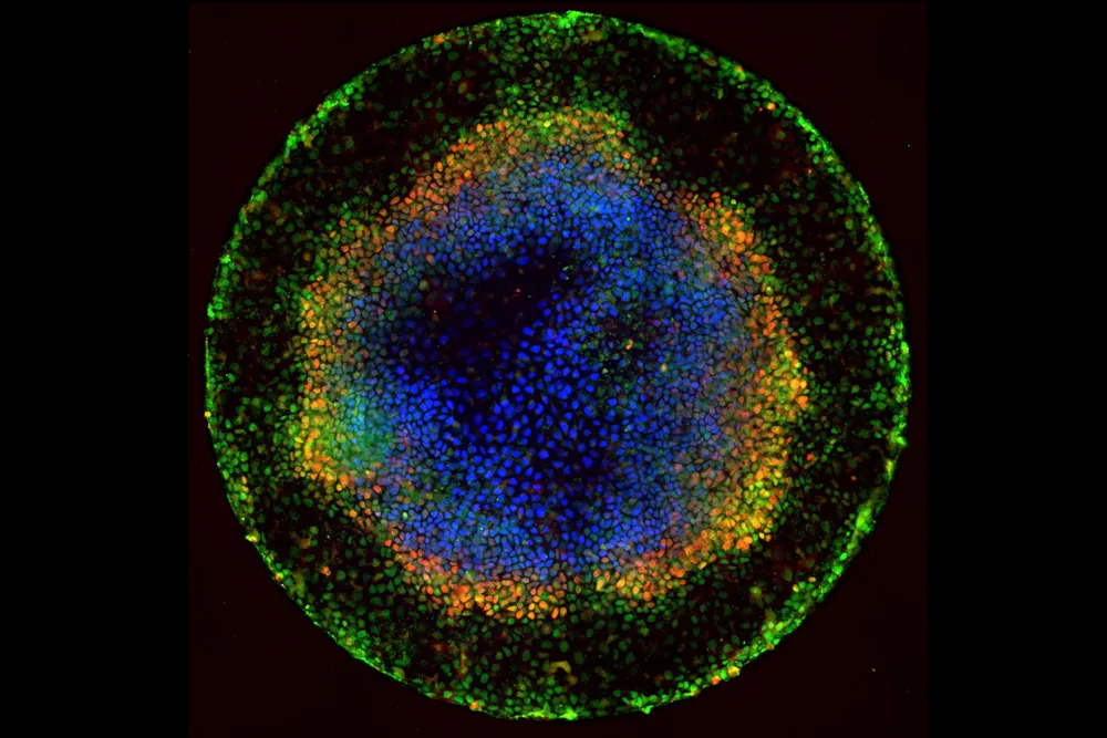
Each fluorescent point of light making up the multicolored rings in this image is an individual human embryonic cell in the early stages of development. These cells are the descendants of human embryonic stem (ES) cells, which have the potential to become any of roughly 200 cell types in the human body.
During normal embryonic development, ES cell descendants specialize, migrate and organize into distinct layers in a process called gastrulation. These layers then shape and fold into structures that give rise to different tissues, organs and limbs. Scientists seeking to understand the molecular cues responsible for early embryonic patterning have focused on finding the right mix of chemical signals, called growth factors, that would allow them to replicate the process in the lab. But a very different approach by a team of National Institutes of Health-funded developmental biologists and physicists at Rockefeller University indicates that a critical element is geometrical.
“Thanks to our diverse scientific perspectives, we were in a good position to realize that geometry could be an important factor,” says developmental biologist Ali Brivanlou, who led the team with physicist Eric Siggia. A former postdoctoral researcher trained in theoretical physics, Aryeh Warmflash, also played a big role.
The researchers grew colonies of human ES cells in tiny circular patterns printed on glass plates, which kept the cells confined to areas of precisely controlled size and shape. Using customized software and fluorescent tags of different colors, the scientists tracked individual cells under a microscope in real time. When they added a growth factor called BMP-4 to the walled-in stem cells, they saw the cells begin to specialize and form organized patterns just as they would under natural conditions. BMP-4-treated cells that were not confined formed random patterns.
The size of the colonies mattered, too. ES cells confined to circles measuring 1 millimeter across — roughly the size and shape of a week-old human embryo — organized into the three main “germ” layers destined to become different human cell types, plus an outer layer of cells like those that become the placenta. Cells confined to smaller circles formed fewer specialized layers, and those in the smallest circles formed only a single germ layer. From these observations, the team concluded that one key way ES cells know their fate is by calculating their distance from the edge of the colony. With the help of mathematical models, the researchers are now looking into exactly how cells make these measurements.
Their follow-up studies of human ES cells confined to micropatterned rectangles, squares and triangles confirm that “the response of a cell to a given growth factor is as much influenced by the geometry as it is by the growth factor itself,” says Brivanlou.
The team’s work has opened a new window for studying early development. Shedding light on the process could advance efforts aimed at using human stem cells to replace diseased cells and regenerate lost or injured body parts, Brivanlou says. “By simply varying the size and geometry of these circles, it might be possible to coax stem cells into becoming brain cells or heart cells or pancreas cells,” he explains.
Get the world’s most fascinating discoveries delivered straight to your inbox.
No stranger to working across disciplines, Brivanlou co-teaches an innovative architecture course on designing “dynamic buildings” of tomorrow that could morph in response to changing environmental conditions or other circumstances, as biological systems can do. His students spend 2 weeks doing experiments in his lab, he says, “so they can appreciate with their own eyes how nature allows forms to change shape.”
The research reported in this article was funded in part by the National Institutes of Health under grants R01GM101653 and R01HD032105.
This Inside Life Science article was provided to LiveScience in cooperation with the National Institute of General Medical Sciences, part of the National Institutes of Health.
Learn more:
All-In-One Stem Cells Article from Inside the Cell Booklet
Also in this series: