In Photos: Ancient Egyptian Skeleton Reveals Earliest Metastatic Cancer
Get the world’s most fascinating discoveries delivered straight to your inbox.
You are now subscribed
Your newsletter sign-up was successful
Want to add more newsletters?

Delivered Daily
Daily Newsletter
Sign up for the latest discoveries, groundbreaking research and fascinating breakthroughs that impact you and the wider world direct to your inbox.

Once a week
Life's Little Mysteries
Feed your curiosity with an exclusive mystery every week, solved with science and delivered direct to your inbox before it's seen anywhere else.

Once a week
How It Works
Sign up to our free science & technology newsletter for your weekly fix of fascinating articles, quick quizzes, amazing images, and more

Delivered daily
Space.com Newsletter
Breaking space news, the latest updates on rocket launches, skywatching events and more!

Once a month
Watch This Space
Sign up to our monthly entertainment newsletter to keep up with all our coverage of the latest sci-fi and space movies, tv shows, games and books.

Once a week
Night Sky This Week
Discover this week's must-see night sky events, moon phases, and stunning astrophotos. Sign up for our skywatching newsletter and explore the universe with us!
Join the club
Get full access to premium articles, exclusive features and a growing list of member rewards.
Burial Position
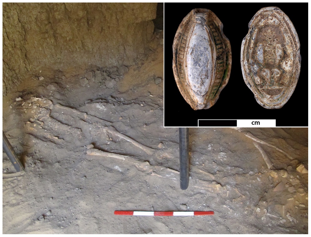
A 3,000-year-old skeleton from a conquered territory of ancient Egypt is now the earliest known complete example of a person with malignant cancer spreading from an organ, findings that could help reveal insights on the evolution of the disease, researchers say. Here, the skeleton (called Skeleton Sk244-8) in its original burial position in a tomb in northern Sudan in northeastern Africa, with a blue-glazed amulet (inset) found buried alongside him; the amulet shows the Egyptian god Bes depicted on the reverse side.
Right Scapula
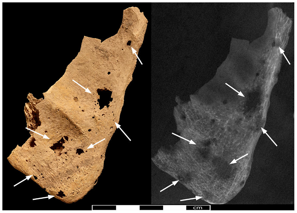
A photo and radiograph of the right scapular blade. The arrows indicate the locations of lytic lesions, which are areas where the bone has been destroyed by a disease process, such as cancer.
Left Clavicle
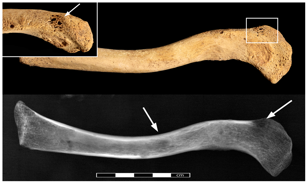
View of the left clavicle with the pathological lesions indicated by arrows. A radiograph of the same bone can be seen on the bottom. At top left, a close-up of the lesion is indicated by the rectangle.
Sternum
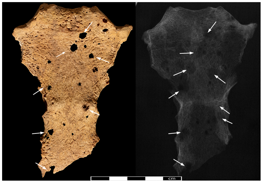
A view of the sternum, with arrows indicating the location of lytic lesions.
Rib
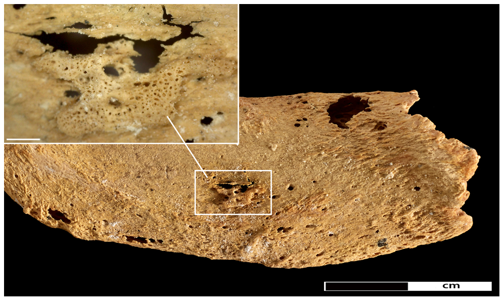
New bone formation on the left first rib. The insert shows a close-up of an area of new bone formation (45× magnification).
Rib
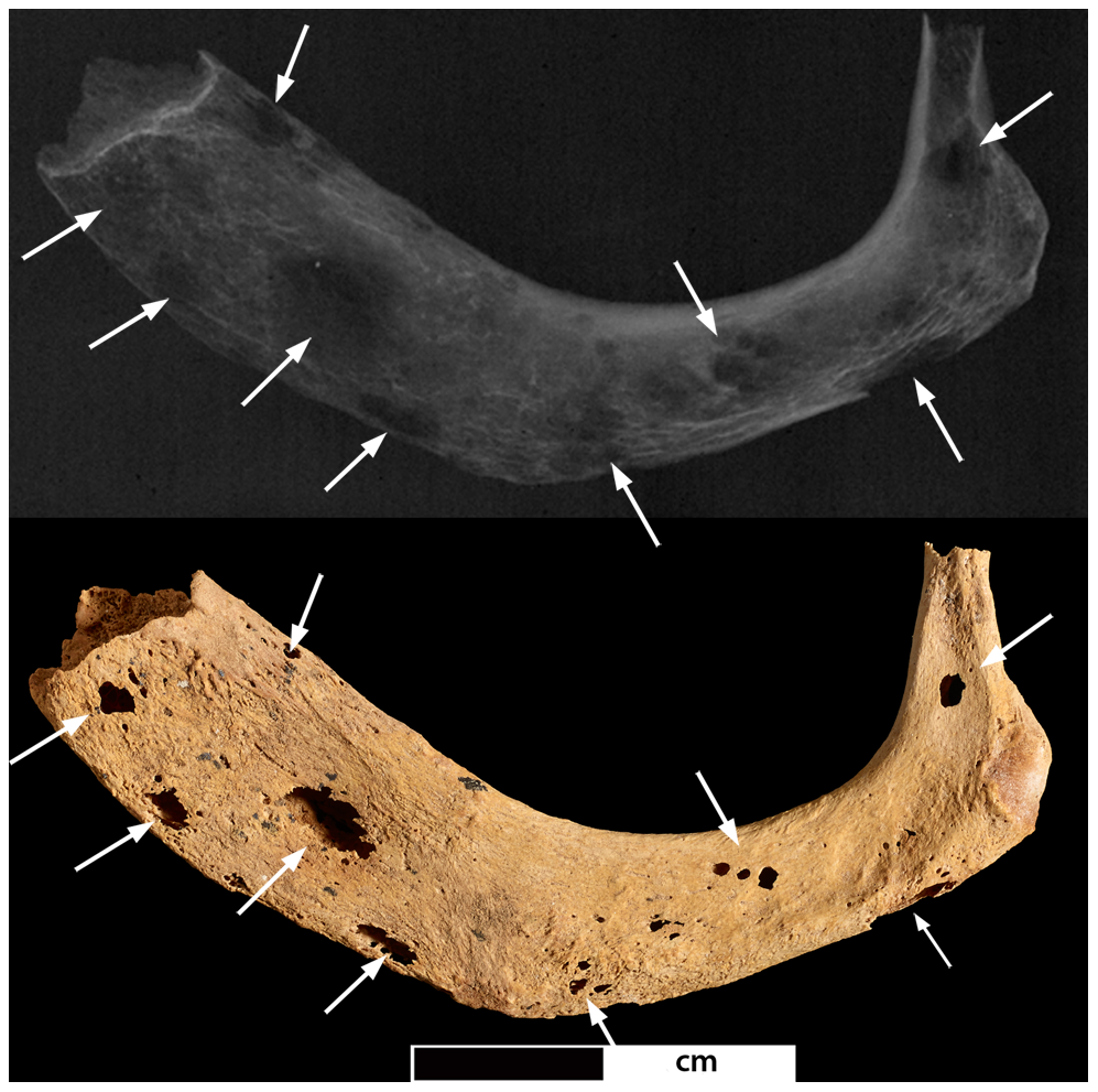
A photo and radiograph of the left first rib. Arrows indicate the locations of lesions.
Thoracic Vertebrae
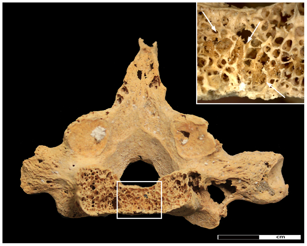
Destructive lesions in the 7th thoracic vertebrae. The arrows indicate regions of new bone formation.
Get the world’s most fascinating discoveries delivered straight to your inbox.
Thoracic Vertebra
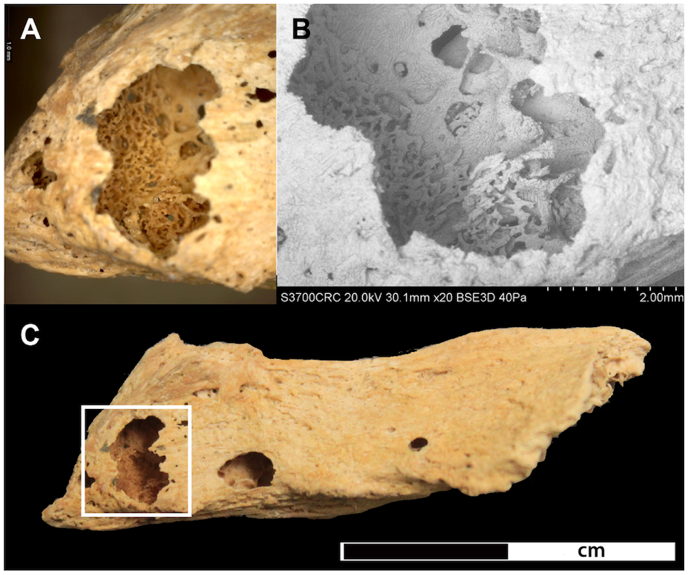
A close-up view of lytic lesions in the 5th thoracic vertebra.
Pelvis
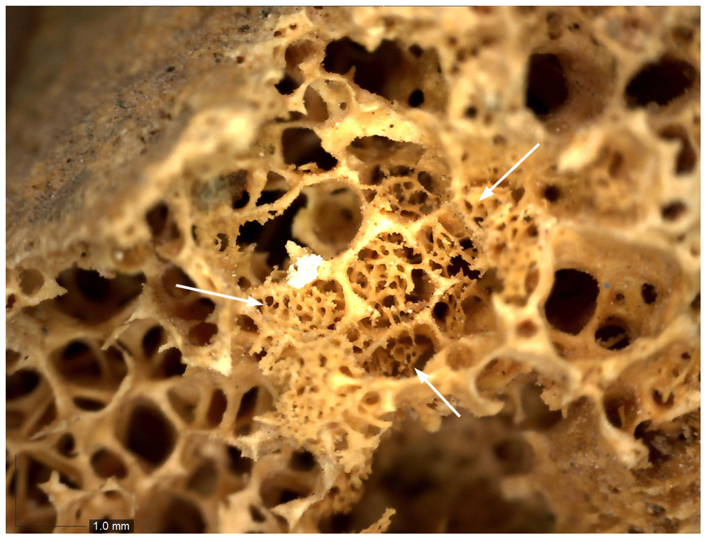
A close-up of new bone formation (indicated by arrows) in a lytic lesion in a part of the pelvis (40x magnified).
Right Femur
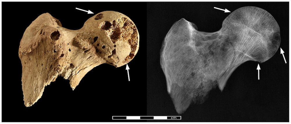
A photo and radiograph of lytic lesions in the right femur.
Preserved Skeleton
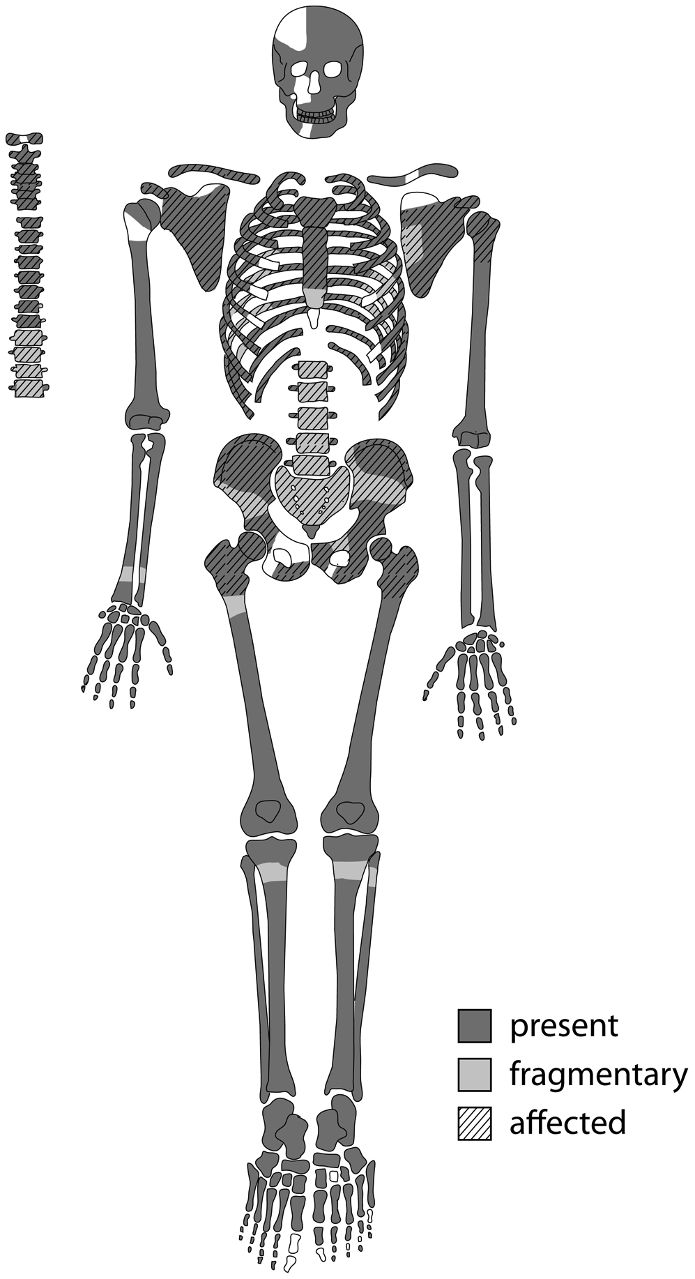
Preserved elements of the ancient Egyptian skeleton and elements affected by pathological changes. Dark areas indicate full preservation, light areas indicate fragmented areas. Hatched areas are the bones affected by lytic lesions.
 Live Science Plus


