
 Live Science Plus


In Photos: A Look Inside an Egyptian Mummy

Suffering Mummy
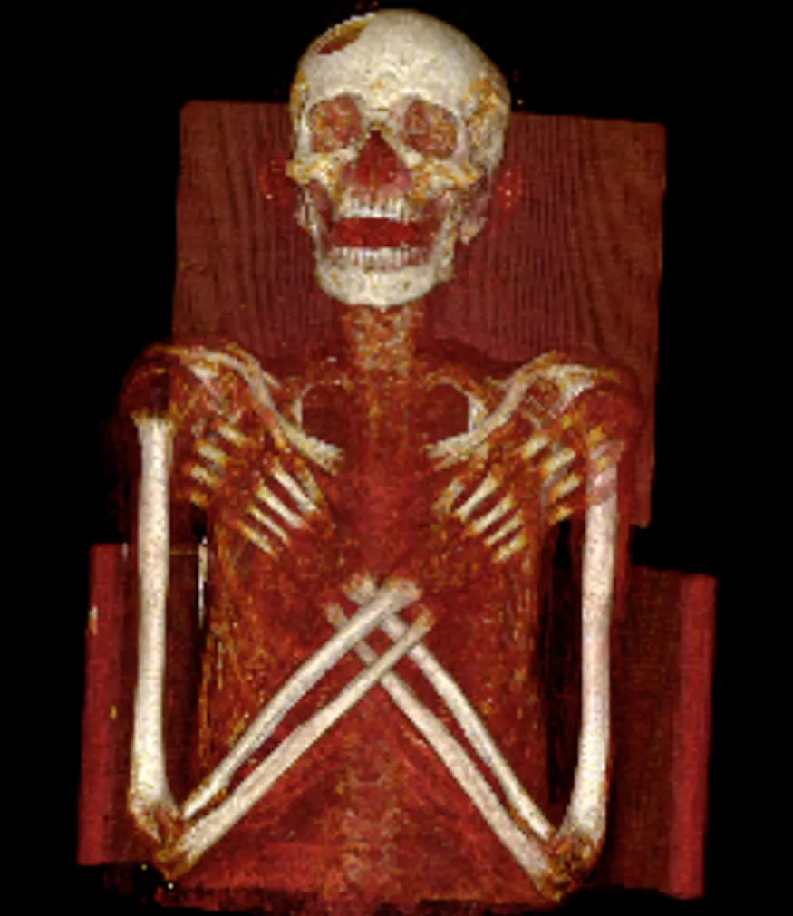
Researchers examined a 2,900-year-old mummy using X-rays, CT and magnetic resonance imaging (MRI) scans. They found that he suffered from Hand-Schuller-Christian’s disease, a very rare condition that left him with lesions in his skull and spine. A large hole on his frontal-parietal bone can be readily seen in this image. His brain appears to have been removed through his nose during the mummification process.
Hole in Head

Normally MRI scans can't be used on mummies, because mummy bodies don't have any water in them. A recently developed technique, however, let the researchers use it to study the mummy of an Egyptian man who likely died in his 20s. In this scan it can be seen that the embalmers filled the back of the mummy's head with a resinlike fluid.
Spinal Dislocation

An image of the mummy's spine in a CT scan. The researchers think spinal dislocation was caused by the embalmers during mummification. Resinlike fluid can be seen in his pelvic area.

Spine Disks

The recently developed MRI technique allowed the researchers to get an up-close look at the inter-vertebral disks of the mummy's spine.
Preserved Penis

Curiously the sarcophagus the mummy was put in belonged to that of a woman named Kareset who lived about 2,300 years ago. The tests showed that the mummy is a man and definitely not Kareset. This image shows the mummy's preserved penis on a CT scan.
Pelvic Picture

An image of the mummy's pelvic area using the new MRI technique. The researchers think the man also suffered from a type of diabetes that would have left his kidneys unable to conserve water. The result would have been that he was thirsty, hungry and urinating all the time.
Bone Replacement

Hand-Schuller-Christian disease causes so-called Langerhans cells, a type of immune cell found in the skin, to multiply rapidly. These cells tend to replace the normal structure of the bone and other soft tissue in the body. Shown here, an MRI scan of the mummy's knees.
