Stunning Photos: Microscopic Images As Art
Bioglyphs Secrets Unveiled
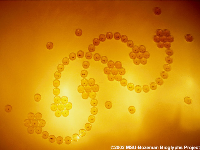
With lights turned on, the secret behind Bioglyphs paintings is revealed -- petri dishes coated with agar support colonies of bioluminescent bacteria. Montana State University-Bozeman School of Art student Angela Bowlds created this piece. Bioglyphs--an exhibition of living bioluminescent paintings--brings science and art together in the form of a collaborative project involving students from the MSU School of Art, and science and engineering students from MSU’s Center for Biofilm Engineering (CBE). For more information about the project, visit the Bioglyphs website
Arch of Bioluminescence
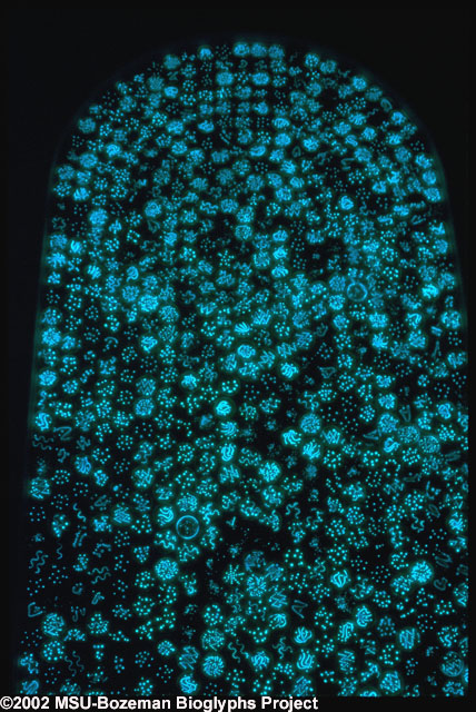
This arch was composed with petri dishes “painted” with bioluminescent bacteria. The piece--approximately 9 feet high by 5 feet wide--was installed in December 2002 at the O’Malley Library, Manhattan College, Riverdale, NY. The painting was created as part of the Bioglyphs project, composed of participants from Montana State University-Bozeman’s Center for Biofilm Engineering (CBE) and School of Art, in collaboration with environmental engineering students of Dr. Robert Sharp at Manhattan College. For more information about the project, visit the Bioglyphs website
Biogeochemical Educational Experiences in South Africa
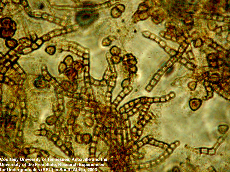
Microscopic photo of metal-oxidizing bacteria found in biofilm samples taken from a South African gold mine. Samples were taken as part of the University of Tennessee's (UT) Biogeochemical Educational Experiences - South Africa (BEE-SA)—a Research Experiences for Undergraduates (REU) program. BEE-SA participants collected fissure water samples from South African gold mines as part of their research. South African mines, particularly the deep gold mines, have been selected for study because they provide relatively easy access to deep fissure waters and the rocks that host them. Since these mines are some of the deepest excavations in the world, they increase the possibility of uncontaminated studies of earlier evolution.
Periphyton Community
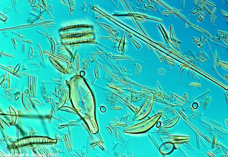
The periphyton community (sessile organisms that live attached to surfaces projecting from the bottom of freshwater aquatic environments) in Lake Hovsgol contains hundreds of diatom species. Diatoms are a large group of microscopic algae that grow as single cells or small colonies. This sample was taken as part of a Mongolian-American international partnership to survey the diatom flora of Hovsgol National Park in north-central Mongolia.
Microscopic Image of Plankton
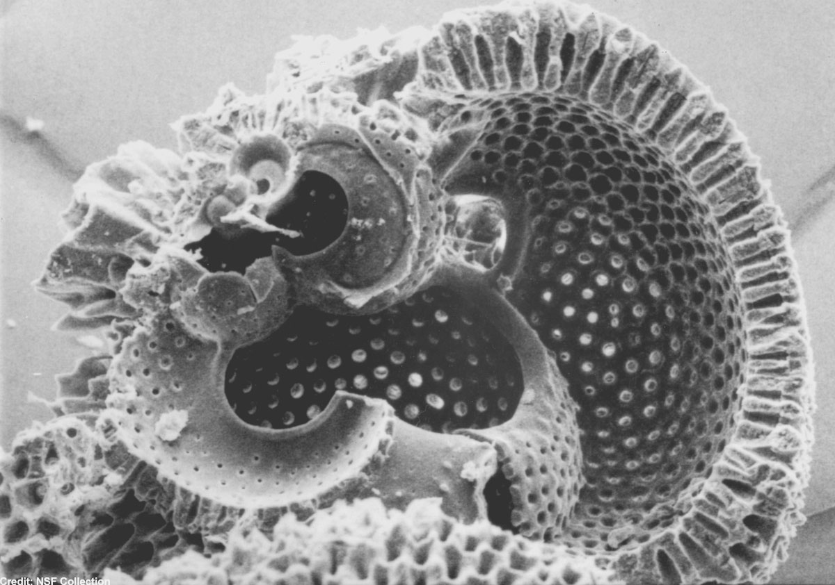
A microscopic image of plankton.
Diatom Species Cymbella stuxbergii
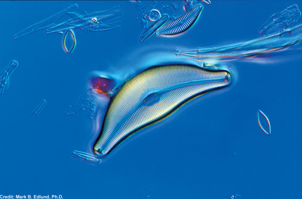
A Cymbella stuxbergii species of diatom. Diatoms are a large group of microscopic algae that grow as single cells or small colonies. This sample was taken as part of a Mongolian-American international partnership to survey the diatom flora of Hovsgol National Park in north-central Mongolia.
Diatom Species Cyclotella ocellata
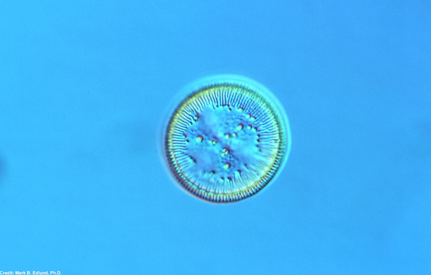
The Cyclotella ocellata species of diatom pictured here is the most common planktonic diatom in Lake Hovsgol, Mongolia. Diatoms are a large group of microscopic algae that grow as single cells or small colonies. This sample was taken as part of a Mongolian-American international partnership to survey the diatom flora of Hovsgol National Park in north-central Mongolia.
Get the world’s most fascinating discoveries delivered straight to your inbox.
Diatom Species Aneumastus
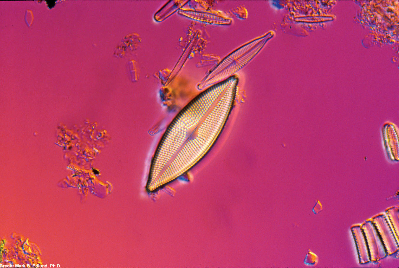
An Aneumastus species of diatom. Diatoms are a large group of microscopic algae that grow as single cells or small colonies. This sample was taken as part of a Mongolian-American international partnership to survey the diatom flora of Hovsgol National Park in north-central Mongolia.
Cholera-Carrying Copepod
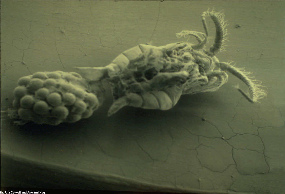
A microscopic view of a female, cholera-carrying copepod.
Name Etched on Human Hair
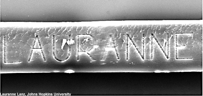
A scanning electron microscope image reveals the word "Lauranne" etched onto a strand of human hair. Ms. Lauranne Lanz, a high school student who participated in a month-long summer internship in July 2002 at the NSF-supported Materials Research Science and Engineering Center (MRSEC) at Johns Hopkins University, performed the etching.
 Live Science Plus


