 Live Science Plus


Small Stature
Check out the following images showing how scientists have been digging up the past lives of these hobbits.
Flores Cave

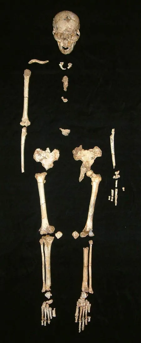
The specimen, along with fossils of various animals, was unearthed in the Liang Bua cave on the island.
Article continues belowHomo erectus
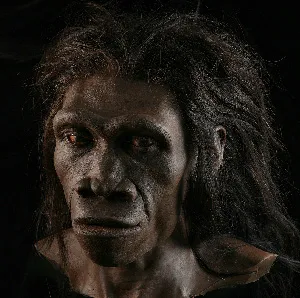
Some scientists named the specimen Homo floresiensis, a dwarfed offshoot of Homo erectus (shown here in artist depiction), a human ancestor that lived as far back as 1.8 million years ago. However, critics remained convinced the specimen was that of a human with microencephalia, a pathological condition characterized by a small head, short stature and varying degrees of mental retardation.
Hobbit Skull

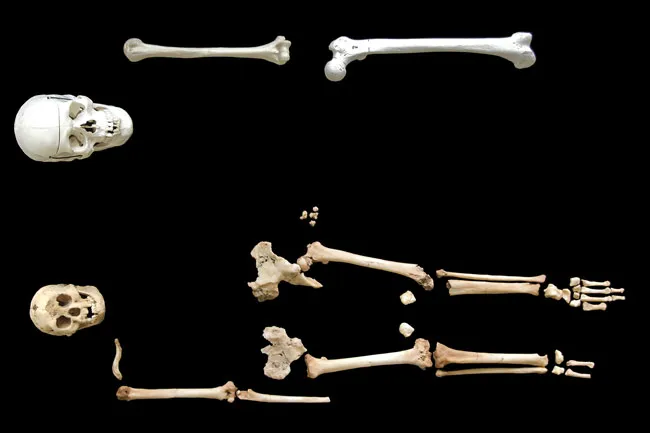
To look into microencephaly, a team of scientists led by Dean Falk, a paleoneurologist at Florida State University, compared computer-generated three-dimensional reconstructions, called "endocasts," of brains from nine microcephalic modern humans with those of 10 normal modern-human brains. They found two ratios created using different skull measurements could accurately distinguish the normal humans (skull, right) from the microcephalics. When Falk's team applied this classification system to a virtual endocast the Hobbit's skull (left), they found its features more closely resembled that of a normal human than a microcephalic.
In the Wrist
In 2007, work by Matthew Tocheri, an anthropologist at the National Museum of Natural History in Washington, D.C., and colleagues found the female Hobbit's wrist bones matched, in shape and orientation, those of non-human apes; they looked much different from the wrist bones of Neanderthals (Homo neanderthalensis) and modern humans, also pointing to a new species.
Long Feet

A detailed analysis of the feet of Homo floresiensis reveals the small-statured hominids, though bipedal, had feet that were so primitive their gait was not efficient; essentially, they couldn't hustle, suggests the 2009 study.
Jawbone Added to 'Hobbit' Evidence

Professor Chris Stringer, Head of Human Origins at London's Natural History Museum, holds a cast taken from a skull that is said to be that of a new species in the evolution of man named 'Homo Floresiensis', during a news conference in London, Wednesday Oct. 27, 2004.
Eaten by Storks?
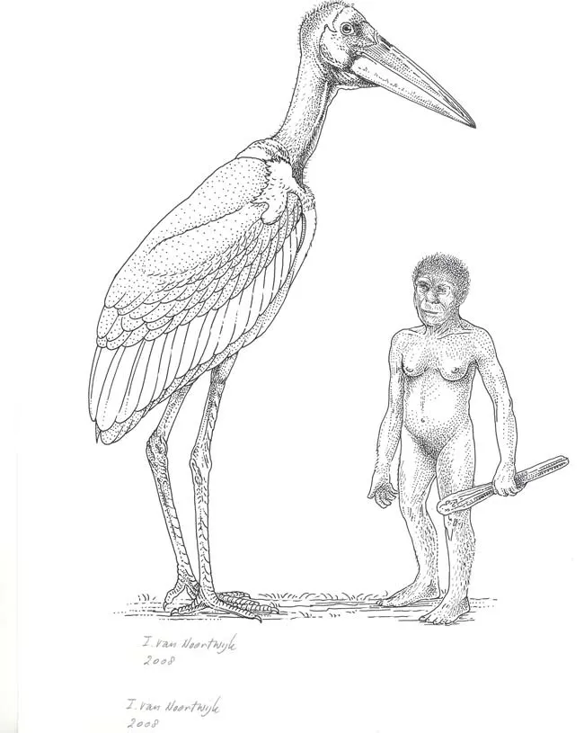
In the Liang Bua cave where the Hobbit remains were found, scientists also discovered a large number of bird fossils, including 20,000- to 50,000-year-old wing and leg bones from what appears to have been a stork that stood nearly 6 feet (1.8 meters) tall. The researchers speculated the extinct predatory stork may have fed on fishes, lizards and birds, and possibly in principle even small, juvenile hobbits, though they note no evidence for such hominid-munching.
Hunting Pygmy Elephants

Her stature combined with evidence from other fossils found at the site paint a picture of a diminutive bipedal individual who used stone tools and fire while hunting the island's pygmy elephants, Komodo dragons and giant rats.
Revealing a Face

In more recent research, anthropologist Susan Hayes, a senior research fellow at University of Wollongong, New South Wales, fleshed out the face of the female Hobbit. To do so, Hayes uploaded information from 3D imaging scans of the skull into a computer graphic program and also looked at portraits by paleo-artists of the Hobbit, finding these earlier interpretations were skewed toward monkey features; her examination, meanwhile, suggested modern features were more accurate.
Facial Features
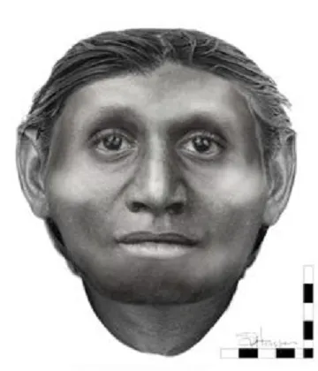
"She's not what you'd call pretty, but she is definitely distinctive," Hayes said. The female doesn't have feminine-looking big eyes and she's lacking much of a forehead.

