
 Live Science Plus


Small World: Gallery of Microscopic Beauty

Kangaroo Rat Kidneys

Keratin filaments in the cytoplasm and nucleus of rat kidney cells help the cells maintain their shape in this Nikon Small World image by Harvard's Lynne Chang. The annual photograph competition highlights microscopic images, and winners will be announced in October. [Gallery of Last Year's Winners]
Tiny Trap
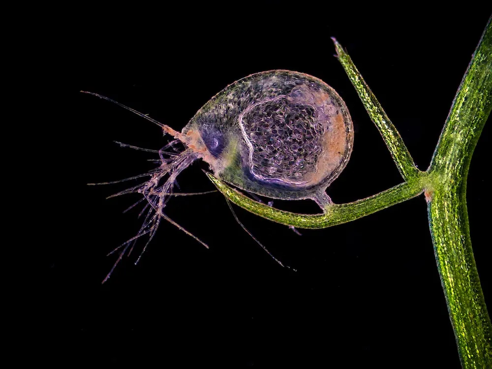
Jose R. Almodovar of the UPR Mayaguez Campus took this photo of a bladderwort bladder for the 2011 Nikon Small World photography contest. Bladderworts are carnivorous plants that trap tiny organisms in their bladders for digestion.
Silver Wattle Anther

Dr. Marta Guervos of the University of Oviedo in Asturias, Spain zoomed in on the anther of a silver wattle or mimosa tree for the 2011 Nikon Small World photography competition.

Red Velvet

Dr. David Maitland of Feltwell, UK gets very close to a red velvet mite, a tiny arachnid, for his Nikon Small World entry.
Earworms

Parasitic filaria worms (in red) inside the lymphatic cells of a mouse's ear, taken for the 2011 Nikon Small World competition by Dr. Witold Kilarski of the EPFL-Laboratory of Lymphatic and Cancer in Lausanne, Switzerland. The worms are a parasitic nematode. Some species are known to infect humans.
Cow Cells

The University of San Francisco's Dr. Torsten Wittmann captured this shot of bovine pulmonary artery cells stained so that actin, mitochondria and DNA appear in yellow and blue.
For more photos, and to vote on your favorite, visit www.nikonsmallworld.com.
Get the world’s most fascinating discoveries delivered straight to your inbox.

Stephanie Pappas is a contributing writer for Live Science, covering topics ranging from geoscience to archaeology to the human brain and behavior. She was previously a senior writer for Live Science but is now a freelancer based in Denver, Colorado, and regularly contributes to Scientific American and The Monitor, the monthly magazine of the American Psychological Association. Stephanie received a bachelor's degree in psychology from the University of South Carolina and a graduate certificate in science communication from the University of California, Santa Cruz.