1st Color X-Rays of Human Body Are Bloody Amazing
Stunning new color X-ray images, from a company called Mars Bioimaging, in New Zealand, seem to make flesh and bone translucent and hyperreal.
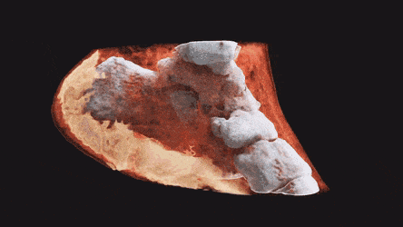
The gif above shows one of the company's strange and fascinating images: a slice of human ankle, with off-white, rugged bones, bloody-looking muscle tissue and a pad of fat smeared protectively under the heel with a whipped-cream texture.
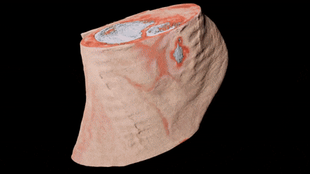
This image shows a wrist with more muscle, less visible bone, almost no fat and a clearly-articulated watch:

It's important to note that these aren't "true-color" X-ray scans as most people would commonly understand the term. As the inventors of the sensor that was used to make these images described in a 2015 paper in the journal IEEE Transactions on Medical Imaging and on the company's website, the colors in these images are applied based on the computer's detection of different wavelengths of X-rays passing through different substances. There are, however, no "true" red X-rays or "true" white X-rays; the device's programmers assign different colors to different detected body parts. (What human brains interpret as color comes from different wavelengths of light in the visual spectrum bouncing off objects. Visible light is also a form of electromagnetic radiation but is lower-energy than X-ray light.)
To successfully distinguish muscle, fat and bone, Mars Bioimaging developed sensors that could fit inside computed tomography (CT) scanners (circular X-ray devices that produce three-dimensional X-ray images) and produce very detailed information about the wavelengths of individual X-ray photons that pass through and bounce off human tissue. By sensing the wavelengths that disappear after passing through a particular bit of tissue, the device makes a judgement about what chemicals make up that tissue and uses that information to figure out what sort of tissue it was. The photon-counting technology, the company says in its marketing materials, was originally developed as part of its founders' work with CERN, the European Organization for Nuclear Research, which operates the world's largest atom smasher.
By matching those scans with details about how different chemical compounds interact with X-ray light, they were able to distinguish different compounds in X-ray scans, the researchers wrote in the 2015 study. To produce these new grody, gorgeous color images of living tissue, they simply tasked the computer with painting the different compounds of fat, bone and muscle different colors.
The benefit for researchers, the company claims in its marketing materials, isn't so much the fascinating visuals (though that's a plus) as it is the wealth of precise chemical data on objects in the scanner. The careful, multilayered tissue scans, they write, will enable new precision in medical research.
Originally published on Live Science.