World of Intricate Muscles Revealed Inside Velvet Worm's Wee Leg
For decades, X-ray computer tomography (CT) scanning has enabled scientists to noninvasively examine the insides of organisms and objects, and model them in 3D. But the technology only worked on subjects that were larger than 500 nanometers (a nanometer is 1-billionth of a meter, or 400-billionths of an inch).
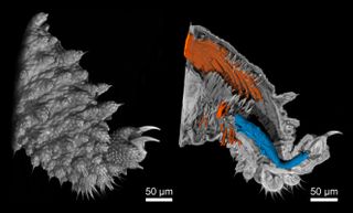
Recently, scientists developed a tabletop Nano-CT system capable of capturing images in 3D at an unprecedentedly small scale — 100 nanometers. Its limits were recently tested on a velvet worm’s minuscule legs, which measure a mere 0.02 inches (0.4 millimeters) long, and this novel technology successfully visualized individual muscle fibers inside the worm's leg, the researchers reported in a new study. [Images: Tiny Life Revealed in Stunning Microscope Photos]
When an object is CT-scanned, multiple X-ray images are taken from many angles, creating cross-sectional views of the object's internal structure. Using computer processing, these individual image "slices" are then combined to rebuild the interior of the image in 3D, according to the Mayo Clinic.
Nano-CT uses nanotubes to tightly focus X-rays and visualize much smaller objects at higher resolution than had been possible with CT scans until now. As the process creates a digital 3D model of the object from a single scan, it is cheaper and less time-consuming to use than other high-resolution imaging methods that can only capture 2D images on a single plane, such as scanning electron microscopy (SEM) and confocal laser scanning microscopy (CLSM), the researchers explained in the study.

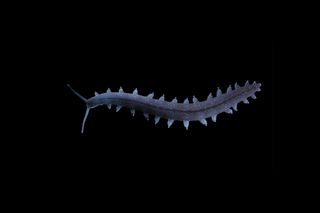
The scientists tested the system by looking inside the legs of the tiny velvet worms —soft-bodied animals that resemble worms with multiple sets of limbs. They are part of the group panarthropoda, which includes arthropods and tardigrades. The scans revealed that the worms' feet contained circular muscles, which had been hinted at in prior studies but were not meticulously described, the scientists reported.
"Our data confirm the existence of these muscles and reveal details of their position, arrangement and size," the researchers wrote. And the characteristics of the muscles suggest that they are used to extend claws in the feet, but the shape and function of most of the velvet worm's muscles are still unknown, according to the study.

Velvet worms are an ancient lineage that has changed little in 500 million years, and their closest relationships on the tree of life are still being debated, the scientists wrote in the study. Internal examination of their delicate limb structures could offer scientists new insights into the animals' locomotion, and may help researchers puzzle out how segmented limbs in arthropods evolved, study co-author Georg Mayer, head of the Department of Zoology at the University of Kassel, said in a statement.
Sign up for the Live Science daily newsletter now
Get the world’s most fascinating discoveries delivered straight to your inbox.
There could also be biomedical applications for this technology, according to Franz Pfeiffer, a professor of biomedical physics at the Technical University of Munich (TUM) and a fellow at the TUM Institute for Advanced Study (TUM-IAS).
"We will be able to examine tissue samples to clarify whether or not a tumor is malignant," Pfeiffer explained in the statement.
"A non-destructive and three-dimensional image of the tissue with a resolution like that of the nano-CT can also provide new insights into the microscopic development of widespread illnesses such as cancer," Pfeiffer said.
The findings were published online Nov. 21 in the journal Proceedings of the National Academy of Sciences.
Original article on Live Science.

Mindy Weisberger is an editor at Scholastic and a former Live Science channel editor and senior writer. She has reported on general science, covering climate change, paleontology, biology, and space. Mindy studied film at Columbia University; prior to Live Science she produced, wrote and directed media for the American Museum of Natural History in New York City. Her videos about dinosaurs, astrophysics, biodiversity and evolution appear in museums and science centers worldwide, earning awards such as the CINE Golden Eagle and the Communicator Award of Excellence. Her writing has also appeared in Scientific American, The Washington Post and How It Works Magazine.
Most Popular


