Images: Tiny Life Revealed in Stunning Microscope Photos
Alien attack
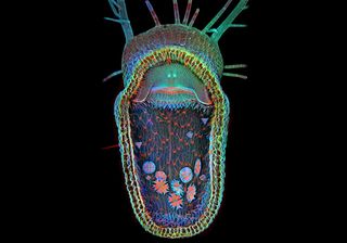
Celebrating its 10th anniversary, the 2013 Olympus BioScapes Digital Imaging Competition honors light-microscope images and movies of humans, plants and animals subjects. Here, the first-place winner reveals the gaping trap of the floating carnivorous plant, a humped bladderwort (Utricularia gibba), taken by Igor Siwanowicz, a neurobiologist at the Howard Hughes Medical Institute's Janelia Farm Research Campus.
Here's a look at the other nine Bioscapes winners.
See no evil
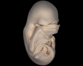
A lateral view of a black mastiff bat embryo (Molossus rufus), at the "Peek-a-boo" stage when its wings have grown to cover its eyes. As development progresses, their fingers grow longer and form the maneuverable struts of their wings, supporting the membrane between their fingers. Stereo microscopy. Second Prize, 2013 Olympus BioScapes Digital Imaging Competition®. www.OlympusBioScapes.com
An intriguing pattern
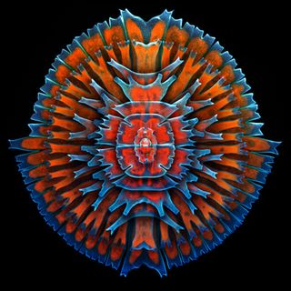
A composite image showing a collection of single-cell fresh water algae, desmids. Desmids exhibit a vast diversity of sizes from 10 microns or smaller to 0.3mm or more. The red in the image comes from the innate fluorescence of chlorophyll. Organisms include (concentric from the outermost in): Micrasterias rotata, Micrasterias sp., M. furcata, M. americana, 2x M. truncata, Euastrum sp. and Cosmarium sp. Confocal imaging, 400x. Third Prize, 2013 Olympus BioScapes Digital Imaging Competition®. www.OlympusBioScapes.com
A start of beauty
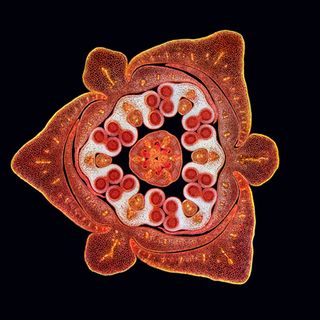
Stained transverse section of a lily flower bud. Darkfield illumination, stitched images. Fourth Prize, 2013 Olympus BioScapes Digital Imaging Competition®. www.OlympusBioScapes.com
A colorful shot
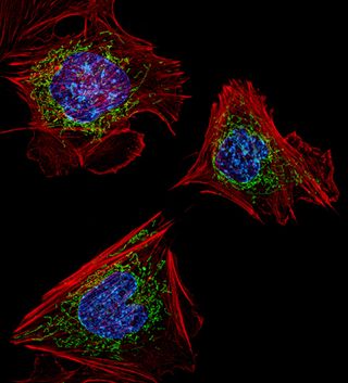
Mouse embryonic fibroblasts showing the actin filaments (red) and DNA (blue). The image also shows the insides of mitochondria, which were visualized by expressing a green fluorescent protein (GFP) fused to a mitochondrial localization sequence. Structured illumination microscopy (SIM) fluorescence; image acquired with a 60x objective. Fifth Prize, 2013 Olympus BioScapes Digital Imaging Competition®. www.OlympusBioScapes.com
Buggie brothers
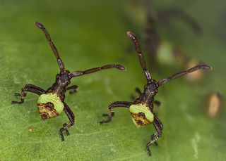
"Brother bugs." Gonocerus acuteangulatus, two hours old. 3mm in size. Sixth Prize, 2013 Olympus BioScapes Digital Imaging Competition®. www.OlympusBioScapes.com
Glassworm
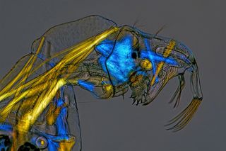
Phantom midge larva (Chaoborus) "Glassworm." Birefringent musculature that is usually clear and colorless is made visible here by specialized illumination. Polarized light, 100X. Seventh Prize, 2013 Olympus BioScapes Digital Imaging Competition®. www.OlympusBioScapes.com
Sign up for the Live Science daily newsletter now
Get the world’s most fascinating discoveries delivered straight to your inbox.
More colors
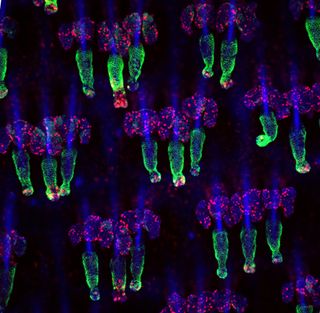
Mouse tail whole mounts stained for the K15 (green) hair follicle stem cell marker as well as Ki67 (red), which marks proliferating cells. Nuclei are marked with DAPI (blue). Technician on the project was Samara Brown. Confocal Z-stack image. Eighth Prize, 2013 Olympus BioScapes Digital Imaging Competition®. www.OlympusBioScapes.com
Head and legs above the rest
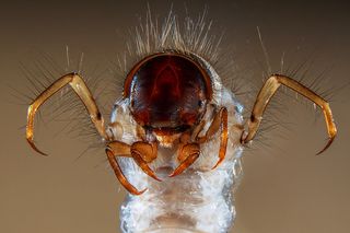
Head and legs of a caddisfly larva: Sericostoma sp., a European and North American genus of insects whose larvae live in fresh water, in gravel, stones or sand. The Sericostoma builds a case (portable tube) of sand grains to protect her flabby body, and eats plant debris and small invertebrates.This larva is a benthic macroinvertebrate that can be used for freshwater biomonitoring; because it is relatively sensitive to organic pollution and dies if water is dirty, it is a good indicator of water quality. Stereo microscopy, 15x. Ninth Prize, 2013 Olympus BioScapes Digital Imaging Competition®. www.OlympusBioScapes.com
Paramecium in action
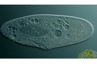
Paramecium, showing contractile vacuole and ciliary motion. Paramecium lives in fresh water. The excess water it takes in via osmosis is collected into two contractile vacuoles, one at each end, which swell and expel water through an opening in the cell membrane. The sweeping motion of the hair-like cilia helps the single-celled organism move. [See the video.] Differential interference contrast, 350x-1000x. Tenth Prize, 2013 Olympus BioScapes Digital Imaging Competition®. www.OlympusBioScapes.com

