Virtual Human Body Changes How Medical Students Learn

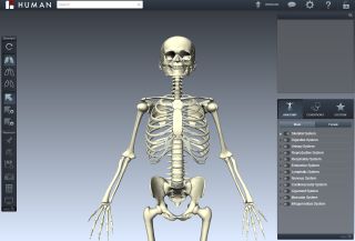
NEW YORK — Today's medical students won't only learn anatomy from a dry, old textbook or a wet, fleshy cadaver. Thanks to NYU School of Medicine and the animation company BioDigital Systems, they can learn using a 3D, virtual, interactive human body.
Its makers call it the BioDigital Human, and LiveScience got a demonstration of the 3D system in action.
Gross anatomy, the study of the human body at the macro scale, remains a pillar of medical education. The dissection of human cadavers in gross anatomy laboratories is considered a rite of passage that most first-year medical students undergo on their paths to becoming doctors.
But whereas human anatomy hasn't changed much over time, technology has evolved dramatically, and it's pumping new life into this age-old subject. Medical schools are now partnering with the computer animation industry to push anatomy education into the 21st century. [In Photos: Explore the BioDigital Human]
Virtual cadavers
Marc Triola, a professor of educational informatics at NYU School of Medicine, and John Qualter, a computer animator and co-founder of Biodigital Systems, unveiled the BioDigital Human at a TEDMED talk in 2012.
The BioDigital Human is not intended to substitute for the gross anatomy lab experience, Qualter said. "I never sought to replace this invaluable learning activity with a computer format, but to emulate it so you could practice it over and over again," Qualter said in the TEDMED talk.
Sign up for the Live Science daily newsletter now
Get the world’s most fascinating discoveries delivered straight to your inbox.
With the click of a mouse, users can navigate a detailed, anatomically accurate model of the human body — bones, muscles, organs and all. They can rotate the body, zoom in, and display, hide or view cross sections of different body systems and structures. Selecting a body part brings up a description that links to its Wikipedia entry (though in theory, it could link to a textbook or any other resource, Qualter said).
The program also contains a wealth of common medical conditions, fleshed out in all their digitally rendered glory. You can even view animations of some of these conditions, such as a heart attack. Or you can view procedures like an appendectomy. And the system is freely available (at least in 2D form) to anyone with an Internet connection. "It's a living digital textbook," Qualter told LiveScience.
Students and faculty members can tag different body parts and create notes using the annotation tool. They can also bookmark these notes to share with others.
From screen to scalpel
At NYU School of Medicine, a life-size version of the BioDigital Human is projected on the wall of the anatomy lab while students are doing real dissections. Students are provided with 3D glasses and iPads (stored in Ziploc bags) so they can magnify and explore the virtual body as they work on a real one.
"It lets the student focus on exactly what it is they need to learn, and it's a terrific supplement to having the cadaver in front of you," said Sally Frenkel, NYU's anatomy course director.
The BioDigital Human was a great resource when Hurricane Sandy hit New York in October 2012. NYU's labs were flooded and unusable, Triola told LiveScience, but the BioDigital Human was fine, safely stored in cloud storage. Students could use it from anywhere — even Starbucks, Triola said.
NYU has also developed a "virtual microscope." It functions like Google Maps, but is designed for human tissue. (It even uses the Google Maps interface.) Faculty at the medical school realized that students often spent vast amounts of time in microscopy labs just trying to get a good slide or focus an image — skills most of them would never need as doctors — so the school replaced real microscopes with the virtual one. The Virtual Microscope is also available for free online.
Students aren't the only ones who can benefit from tools such as the BioDigital Human and the Virtual Microscope, Triola said. Doctors could use both tools to brush up on their knowledge throughout their careers, and patients could use them to learn about their conditions, he said.
Medical technology is advancing rapidly. Qualter and colleagues are currently working on a human body system users could control in a manner similar to the Microsoft Kinect motion sensor. Someday, Qualter said, he envisions some kind of virtual reality system for interacting with a digital body. "But I think that's many years away," he said.
Follow Tanya Lewis on Twitter and Google+. Follow us @livescience, Facebook & Google+. Original article on Live Science.

Most Popular


