How a Jellyfish Protein Transformed Science

These days, scientists can track how cancer cells spread, how HIV infections progress and even which male ends up fertilizing a female fruit fly's egg. These and many other studies that offer insight into human health all benefit from a green, glowing protein first found in a sea creature.
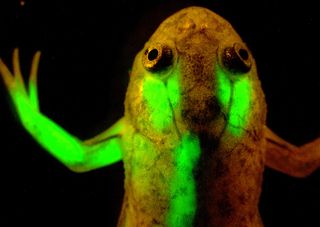
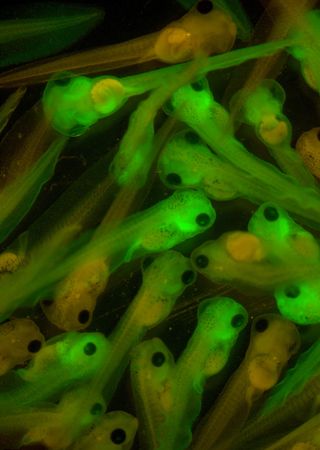
From its humble beginnings in the bodies of a particular species of jellyfish, green fluorescent protein, or GFP for short, has transformed biomedical research. Using a gene that carries instructions to make GFP, scientists can attach harmless glow-in-the-dark tags to selected proteins, either in cells in lab dishes or inside living creatures, to track their activity. It's like shining a flashlight on the inner workings of cells. Flatworms, tadpoles and zebrafish are among the creatures whose parts have been programmed to glow in the name of science.
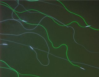
Individual proteins in human cells are too small to see even under a microscope, but the ways they form, fold and interact are essential to life — so catching a glimpse of them in action is invaluable.For example, attaching GFP to insulin-producing cells in the pancreas has helped researchers study how they're made, which could inform new diabetes treatments. Tagging fruit fly sperm with GFP is likewise shedding light on how certain sex cells maximize their chances of being fertilized.
GFP can also be used to study aspects of the intracellular environment, such as a cell's pH or calcium concentration.
Ancient Origins
GFP might sound like a futuristic, sci-fi material, but it has actually been around for more than 160 million years. The protein is naturally expressed in the North American jellyfish Aequorea victoria, and works by absorbing energy from blue light in the environment and emitting a green glow in response. Scientists don't know why these jellyfish evolved their glow, but one hypothesis is that it helps them ward off predators.
Jellyfish aren't the only bioluminescent (making their own glow) creatures on the planet. Differently colored glowing proteins occur naturally in more than a hundred species, including fireflies and corals. Many of these fluorescent proteins are being used to derive new colored tags for research.
Sign up for the Live Science daily newsletter now
Get the world’s most fascinating discoveries delivered straight to your inbox.

GFP remains special, however, because it spontaneously folds into the right shape when it's created, ready to glow without needing help from additional chemicals, enzymes or other substances.
A Prized Protein
GFP research had its time to shine on stage in 2008 when Martin Chalfie, Osamu Shimomura and Roger Tsien received the Nobel Prize in chemistry for their discovery and development of GFP.
Shimomura first extracted the glowing protein from jellyfish in the 1960s and then isolated the gene that carries the instructions to make it. This enabled researchers to introduce the gene into virtually any other animal through breeding or by using techniques such as viruses to deliver the genetic material. As long as an animal has the gene, it can produce GFP — which can be used to light up specific parts of an animal, like muscle cells or brain cell circuits.

Chalfie's work in the early 1990s turned GFP into a powerful tool for studying gene expression, and Tsien's work in subsequent years produced an entire palette of colors that glowed longer and brighter, allowing researchers to study many different molecules at the same time.
The discovery of GFP opened the door to exciting new scientific possibilities, like seeing how specific sets of cells behave during embryonic development, studying how cells die during a process called apoptosis and even finding new ways to turn fluorescence on and off.
Going Beyond GFP
GFP is good for studying cells, but it does have its limitations. In live mammals, its fluorescent colors are often masked because hemoglobinin the blood absorbs the visible light that GFP emits.
A team of researchers at Albert Einstein College of Medicine engineered a new fluorescent probe using a bacterial protein. Dubbed iRFP, the probe emits near-infrared light. Since mammalian tissues are nearly transparent in infrared light, this probe makes it easy to peer inside whole organs and organisms.
So far, iRFP has only been tested on mice. But as a nontoxic, noninvasive method that doesn't involve radiation, iRFP could have great potential for imaging in humans.
Learn more:
- Green Light: profile of Marc Zimmer
- Cool Tools on ChemHealthWeb
- "Green Fluorescent Protein" in 21st Century Genetics
This Inside Life Science article was provided to LiveScience in cooperation with the National Institute of General Medical Sciences, part of the National Institutes of Health.

